The nucleus, shown in Fig 1.1, is a defining feature of eukaryotic cells, enclosed by a specialized membrane system. (a) Describe the structure of the nuclear envelope. [4] (b) Explain the importance of nuclear pores in the function of the nucleus. [3]
The rough endoplasmic reticulum (RER) is a prominent organelle in cells that are highly active in protein synthesis and secretion. (a) Discuss the structural features of rough endoplasmic reticulum and explain how these features facilitate its function in protein synthesis and modification. [6] (b) Draw a labelled diagram of a section of rough endoplasmic reticulum, showing its association with ribosomes and the nuclear envelope. [4]

Fig 1.1 shows an electron micrograph of a bacterial cell. (a) Calculate the actual length of the bacterial cell in micrometers (µm). [3] (b) Explain why it is important to calibrate an eyepiece graticule for each objective lens used with a light microscope. [4]

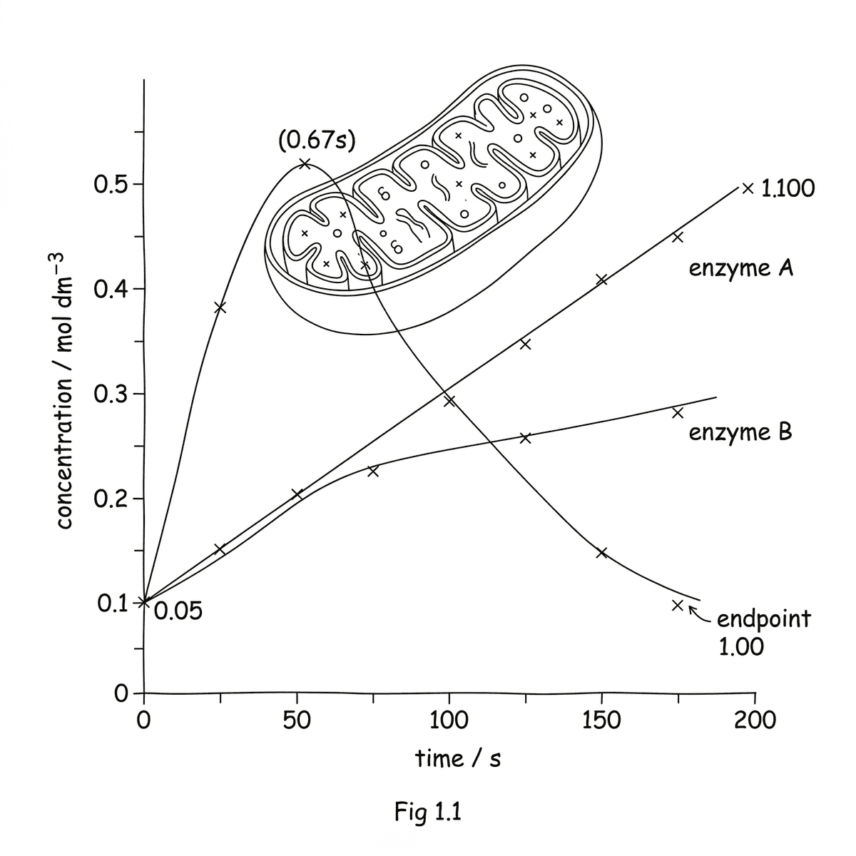
Lysosomal enzymes play a critical role in cellular processes. The graph in Fig 1.1 shows the concentration of lysosomal enzymes within a cell over a period of 10 hours, in response to a specific stimulus. (a) Interpret the graph to describe how the concentration of lysosomal enzymes changes over time in the presence of a specific stimulus. [3] (b) Explain how the observed changes in lysosomal enzyme concentration might contribute to the cell's response to the stimulus. [4]
Light microscopes are essential tools for observing cells and their structures. Accurate measurement of cell size often requires the use of an eyepiece graticule calibrated against a stage micrometer. (a) Draw a simple diagram of a light microscope and label four key components involved in focusing and magnifying the image. [4] (b) An eyepiece graticule has 100 divisions. When calibrated with a stage micrometer, 20 divisions of the eyepiece graticule correspond to 0.1 mm. Calculate the actual size of a cell that measures 60 divisions using this eyepiece graticule. [4]
Electron microscopes are powerful tools used to visualise the intricate structures within cells. (a) Explain why electron microscopes have a higher resolution than light microscopes. [3] (b) An organelle appears 50 mm long in an electron micrograph taken at a magnification of ×250 000. Calculate the actual size of this organelle. Give your answer in micrometers (µm). [4]
The cell surface membrane is a vital component of all living cells, controlling the passage of substances into and out of the cell. (a) Describe the general structure of the cell surface membrane. [3] (b) Explain how the partially permeable nature of the cell surface membrane is crucial for cell function. [5]
Ribosomes are essential organelles found in all living cells, playing a crucial role in protein synthesis. (a) Name the two types of molecules that make up a ribosome. [2] (b) State the primary function of ribosomes in a cell. [2] (c) Identify the difference in size between prokaryotic and eukaryotic ribosomes. [2]
Bacteria possess several distinct genetic elements crucial for their survival and adaptation. (a) Describe the key structural features of a plasmid in a bacterial cell. [4] (b) A plasmid has an actual length of 2.5 µm. If it is observed in an electron micrograph with a measured length of 50 mm, calculate the magnification of the image. [4]
Cells are the basic units of life and can be observed using a light microscope. (a) Identify two organelles that are clearly visible in both plant and animal cells when viewed with a light microscope. [2] (b) State the approximate size range of a typical plant cell when viewed with a light microscope. [2] (c) Describe one difference in appearance between the nucleus of a plant cell and an animal cell under a light microscope. [2]
Plant and animal cells are both eukaryotic cells but exhibit significant structural differences. (a) Compare the structure of a mature plant cell with that of an animal cell as observed using an electron microscope, highlighting three key differences. [6] (b) Discuss how the presence of a cell wall and a large central vacuole in plant cells are advantageous for their functions. [4]
Eukaryotic cells, whether plant or animal, share several common fundamental structures. (a) Draw a diagram of a generalised eukaryotic cell as seen with a light microscope, showing structures common to both plant and animal cells. Label five of these structures. [5] (b) Explain the function of two of the labelled structures identified in (a). [4]
Prokaryotic cells, such as bacteria, possess genetic material in a different arrangement compared to eukaryotic cells. (a) Describe the structure and location of circular DNA in a typical prokaryotic cell. [5] (b) A plasmid has an observed diameter of 0.5 cm in an electron micrograph with a magnification of ×20000. Calculate the actual diameter of the plasmid in micrometres. [3]
Plant cells and bacterial cells both possess a cell wall, but their structures and compositions differ. (a) State two main functions of the cell wall in plant cells. [2] (b) Identify the primary component of the plant cell wall. [2] (c) Give one difference in composition between a bacterial cell wall and a plant cell wall. [2]
Lysosomes play a crucial role in maintaining cellular health and responding to cellular stress. (a) Define the term 'self-digestion' in the context of a cell. [2] (b) State two situations where self-digestion might occur in a eukaryotic cell and identify the organelle primarily responsible. [4]
The endoplasmic reticulum (ER) is a vital network of membranes within eukaryotic cells, playing a crucial role in cellular processes. (a) Identify two distinct types of endoplasmic reticulum found in eukaryotic cells. [2] (b) Outline the general role of the endoplasmic reticulum in the transport of molecules within a cell. [4]
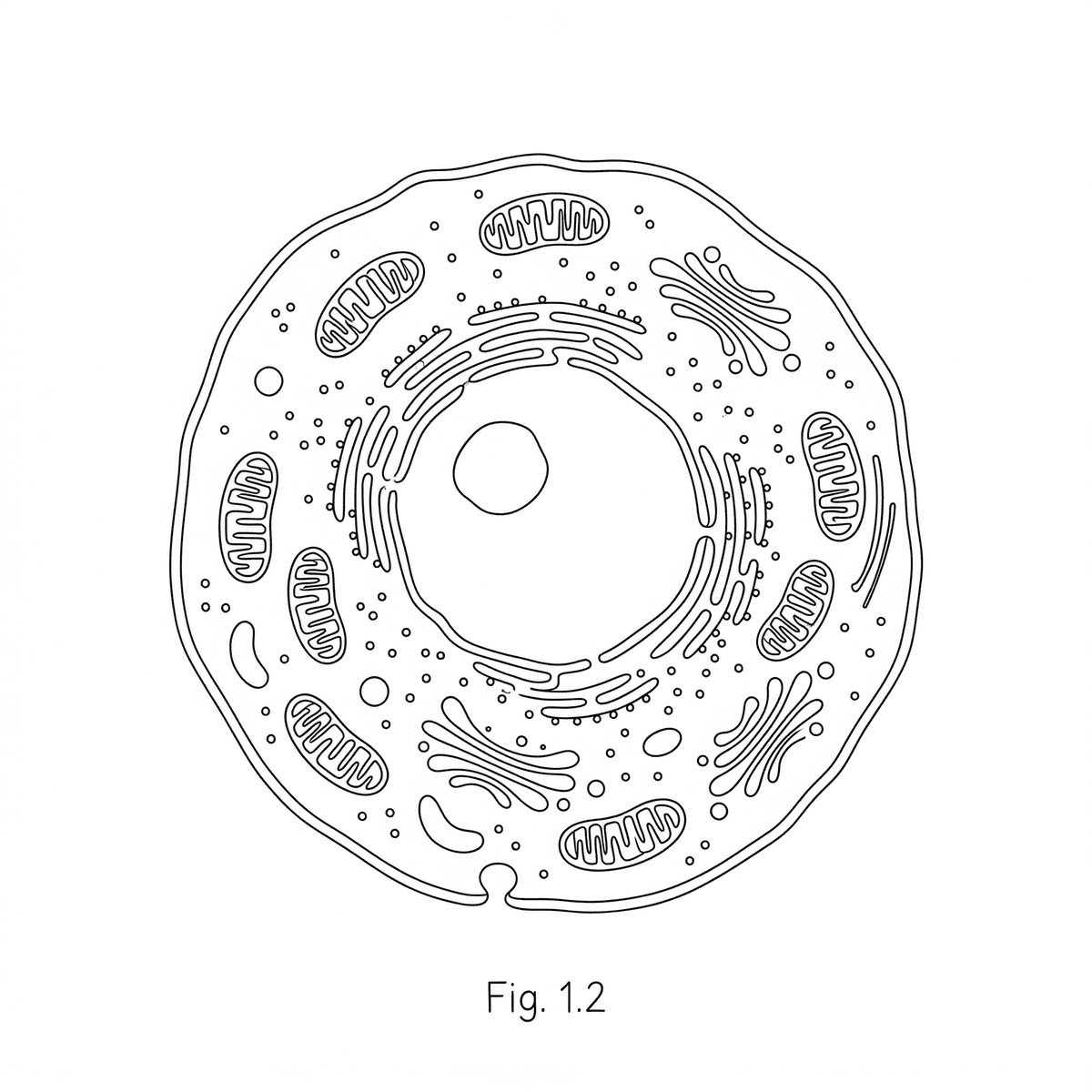
Fig 1.2 shows a transmission electron micrograph (TEM) of a mitochondrion, clearly illustrating its double membrane and the folded inner membrane forming cristae. (a) Measure the observed length of the mitochondrion from Fig 1.2 and use the scale bar to determine its actual length in micrometers (µm). [3] (b) Calculate the approximate surface area of the inner mitochondrial membrane if it were unfolded, assuming the mitochondrion is cylindrical. Use the actual length you calculated in (a) and an estimated actual diameter of 0.5 µm. Assume the cristae increase the surface area by a factor of 5 compared to a smooth inner membrane of the same dimensions. [4] (c) Explain the significance of the large surface area of the inner mitochondrial membrane for its function. [3]

Fig. 1.1 shows an eyepiece graticule scale and a stage micrometer scale used for microscopical measurement. (a) Calculate the calibration of one eyepiece graticule unit in micrometers (µm). (b) A particular cell is measured to be 40 eyepiece graticule units long. Determine the actual length of this cell in micrometers (µm).
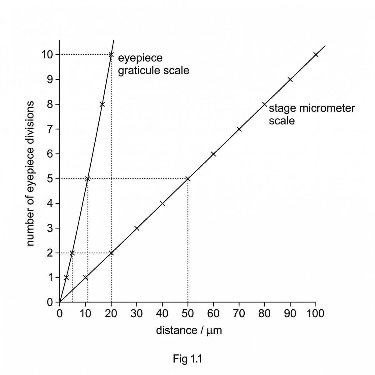
Fig 1.4 shows a diagram of a cilium in transverse section (TS), illustrating its characteristic '9 + 2' arrangement of microtubules and a basal body. A scale bar representing 0.2 µm is provided. (a) Measure the observed diameter of the cilium in transverse section from Fig 1.4 and use the scale bar to determine its actual diameter. (b) Calculate the total number of tubulin dimers present in one cross-section of a cilium, given that each microtubule is made of 13 protofilaments and each protofilament consists of tubulin dimers. Assume one tubulin dimer per protofilament in a cross-section. (c) Explain how the '9+2' arrangement of microtubules contributes to the beating mechanism of cilia.
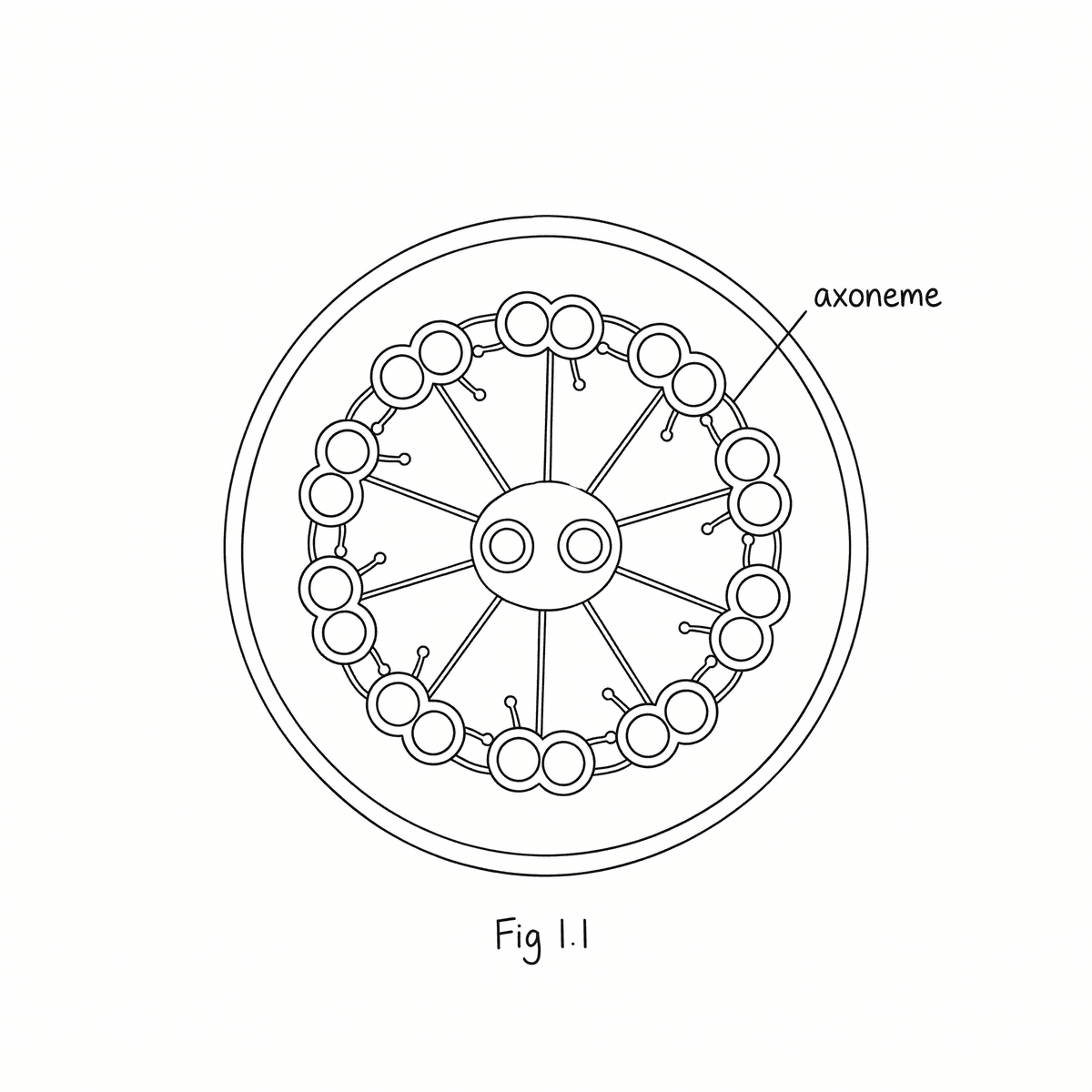
Fig. 1.1 shows a diagram of a mitochondrion, an organelle vital for cellular respiration. (a) Identify the labelled structures X and Y in Fig. 1.1. [2] (b) Explain the significance of the folds of the inner membrane (Y) shown in Fig. 1.1. [3] (c) Draw a simple diagram to show how a molecule of ATP is formed from ADP and phosphate, indicating where the energy is stored. [3]
Chloroplasts are organelles found in plant cells and algal cells, where photosynthesis takes place. Fig. 1.2 shows an electron micrograph of a chloroplast. (a) Identify the structures labelled P and Q in Fig. 1.2. [2] (b) The observed diameter of the chloroplast in Fig. 1.2 is 75 mm. If the actual diameter of the chloroplast is 5 µm, calculate the magnification of the image. Show your working. [4] (c) Explain how the internal structure of a chloroplast is adapted for its function. [4]

Viruses are unique biological entities that blur the line between living and non-living. Their existence highlights fundamental differences in biological organisation. (a) Describe the basic structure of a virus, including the components that may be present. [4] (b) Discuss why viruses are considered non-living and how their mode of replication differs from that of prokaryotic or eukaryotic cells. [6]
Plant cells possess a cell wall, a rigid outer layer that provides structural support. (a) State two functions of the cell wall in plant cells. [2] (b) Describe the structure of a plant cell wall. [4]
Electron microscopy requires specific specimen preparation techniques to ensure high-quality images. (a) State two reasons why specimens must be prepared in a vacuum for electron microscopy. [2] (b) Outline the main steps involved in preparing a biological specimen for viewing with a Transmission Electron Microscope (TEM). [4]
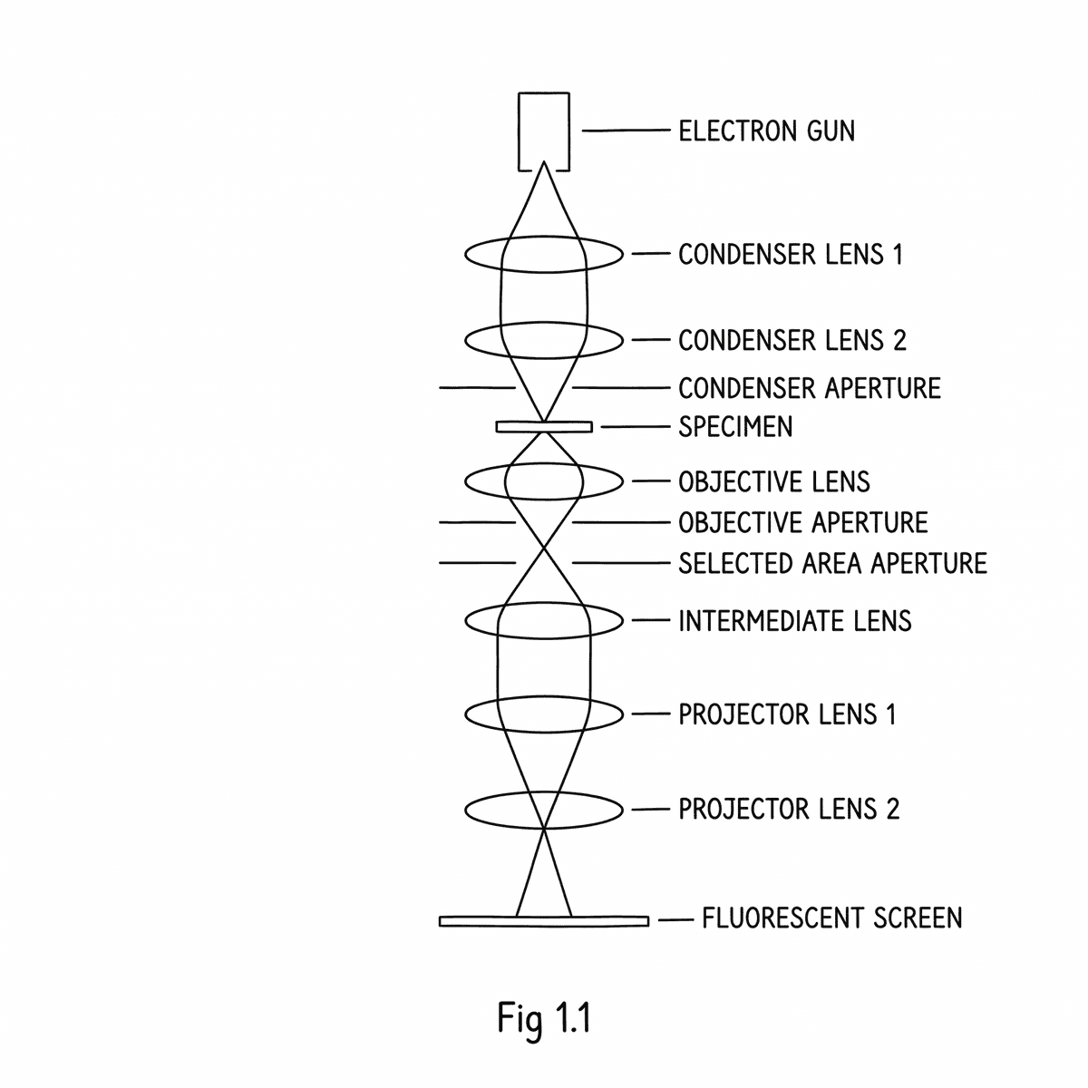
Electron microscopy offers detailed insights into cellular structures, but different types of electron microscopes serve distinct purposes. Fig. 1.1 shows a simplified representation of the beam paths in a Transmission Electron Microscope (TEM) and a Scanning Electron Microscope (SEM). (a) Compare the principles of image formation and the types of images produced by a Transmission Electron Microscope (TEM) and a Scanning Electron Microscope (SEM). [6] (b) Discuss one advantage and one disadvantage of using an SEM compared to a TEM for studying cell surfaces. [4]
The endosymbiont theory proposes that mitochondria and chloroplasts originated from free-living prokaryotic cells that were engulfed by a host cell. (a) Discuss the evidence that supports the endosymbiont theory for the origin of mitochondria. [6] (b) Suggest why the evolution of mitochondria was a significant step in the evolution of eukaryotic life. [4]
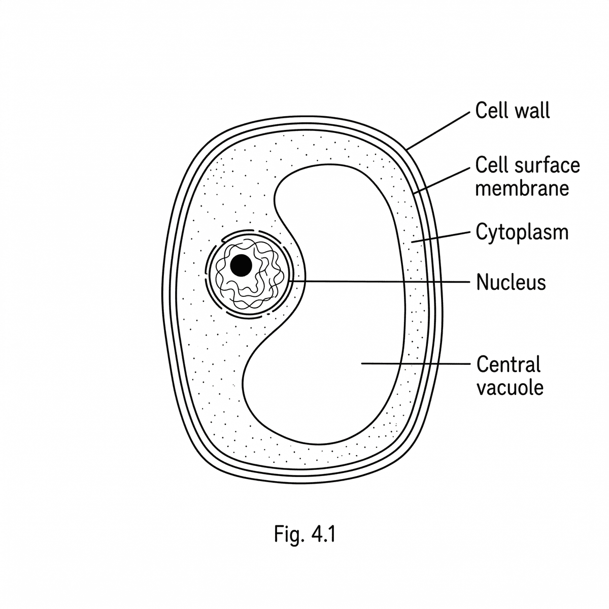
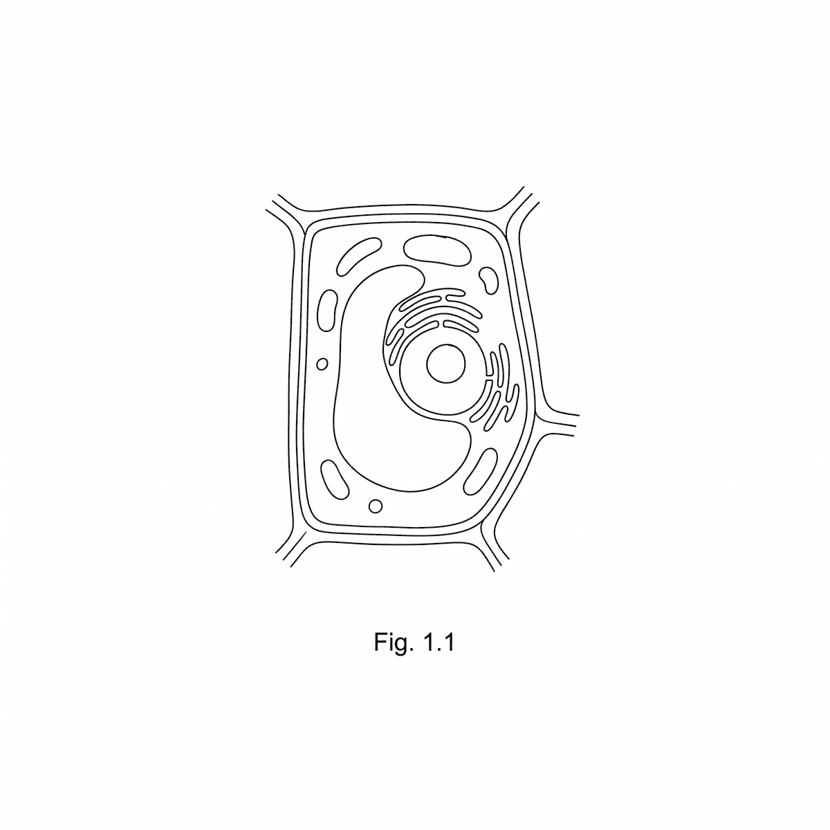
Fig 1.3 shows a drawing of a generalised plant cell as seen with a light microscope. (a) Measure the observed length of the large central vacuole in the plant cell drawing and use the scale bar to determine its actual length. (b) Calculate the approximate volume of the vacuole, assuming it is cylindrical with the measured length and an actual diameter of 15 µm. Give your answer in µm³. (c) Explain how the large central vacuole contributes to the support of the plant cell.
The Golgi apparatus is a vital organelle in eukaryotic cells, involved in modifying, sorting, and packaging proteins and lipids for secretion or delivery to other organelles. (a) Describe the process of protein modification and packaging within the Golgi apparatus. [5] (b) Draw a simple diagram of the Golgi apparatus, labelling the cis face, trans face, and Golgi vesicles. [3]
Lysosomes are important organelles involved in the breakdown of various cellular components. (a) Describe the structure of a lysosome. [3] (b) Explain how lysosomes are involved in the digestion of worn-out organelles. [4]
Cells constantly maintain their internal environment by removing unwanted or damaged components. (a) Name two types of unwanted cell components that might be removed by a lysosome. [2] (b) Outline the general role of hydrolytic enzymes in the breakdown of these components. [3]
Organisms store energy in various forms to meet their metabolic demands. Plants primarily use starch, while animals use glycogen, as their main carbohydrate food reserves. (a) Describe the main difference in the chemical composition of starch and glycogen as food reserves. [4] (b) A plant cell stores starch granules that have an observed diameter of 2.5 cm in an electron micrograph taken at a magnification of ×25000. Calculate the actual diameter of the starch granule in micrometres (µm). [4]
Cell walls are found in various organisms, including plants and bacteria, but their composition and precise functions can differ. (a) Compare the structure and composition of the cell wall in a plant cell with that of a bacterial cell. [6] (b) Explain the importance of plasmodesmata in plant cells. [4]
Fig 1.2 shows an electron micrograph of a plant cell. (a) The observed diameter of a chloroplast within the cell is 45 mm. The scale bar provided on the micrograph is 10 mm long and represents an actual length of 1 µm. Calculate the actual diameter of the chloroplast in micrometres (µm). [3] (b) Determine the magnification of the image if the actual diameter of the nucleus is 15 µm and its observed diameter in the micrograph is 30 mm. [3] (c) Discuss the challenges of accurately measuring the size of organelles from micrographs, considering potential sources of error. [4]
Fig 1.2 shows a transmission electron micrograph (TEM) of a cell surface membrane at very high magnification, revealing its characteristic three-layered appearance. A scale bar is included. (a) Measure the observed thickness of the cell surface membrane from Fig 1.2 and the observed length of the scale bar. [2] (b) Calculate the actual thickness of the cell surface membrane in nanometers. [2]
Bacteria are single-celled prokaryotic organisms that exhibit a unique cellular organisation. (a) Draw a large, labelled diagram of a bacterium, showing its major organelles and structures. [6] (b) Calculate the actual diameter of a bacterium that appears 50 mm long in a micrograph taken at a magnification of ×25 000. Give your answer in micrometres (µm). [4]
Fig 1.4 shows a transmission electron micrograph (TEM) of a nucleus from a bat pancreas cell, clearly depicting the nuclear envelope and several nuclear pores. (a) Measure the observed diameter of a nuclear pore from Fig 1.4. [2] (b) Using the scale bar provided in Fig 1.4, calculate the actual diameter of the nuclear pore in nanometers (nm). [3]
Fig 1.1 shows a bacterial cell viewed under an electron microscope. (a) The observed length of the bacterial cell is 50 mm on the micrograph. The magnification of the micrograph is ×10 000. Calculate the actual length of the bacterial cell in micrometres (µm). [3] (b) Convert the length calculated in (a) into millimetres (mm). [2] (c) Explain why it is important to use appropriate units of measurement when describing cell sizes. [3]
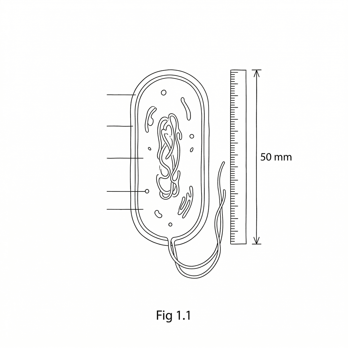
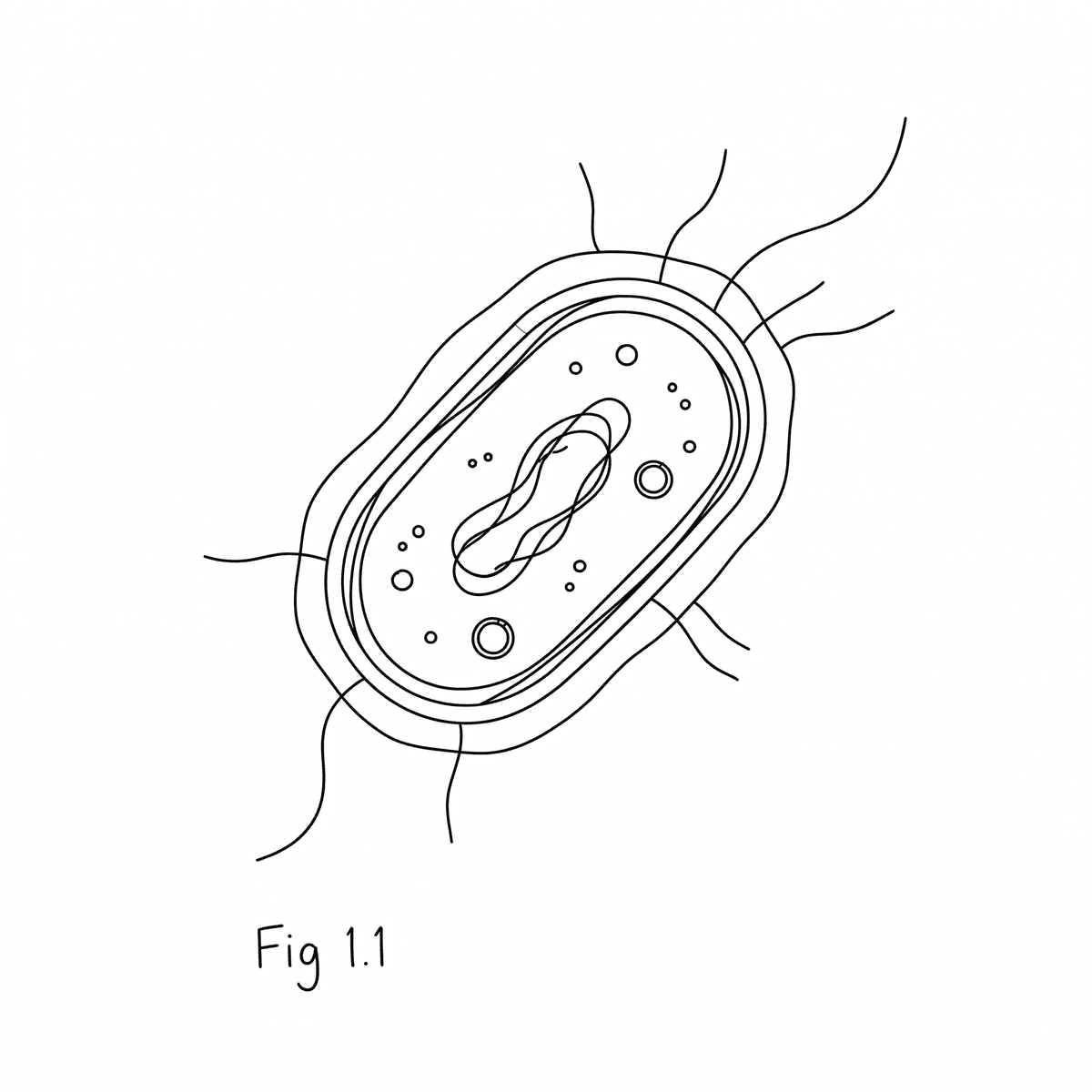
Fig 1.1 shows a simplified diagram of a bacterial cell, highlighting its protective layers. (a) Compare the composition and location of a bacterial capsule with its cell wall. [4] (b) Discuss the advantages a capsule provides to pathogenic bacteria in terms of survival and infection. [6]
Microscopy is a fundamental tool in biology, allowing scientists to observe the intricate structures of cells and tissues. Two key characteristics of a microscope's performance are magnification and resolution. (a) Define the term 'resolution' in microscopy. [2] (b) A student views a plant cell under a light microscope. The observed length of the cell is 48 mm, and its actual size is 120 µm. Calculate the magnification used to view this cell. Show your working. [4] (c) Explain why increasing the magnification beyond a certain point does not necessarily lead to more detail being observed. [2]
Fig 1.1 shows an electron micrograph of a mitochondrion (labelled X) within a eukaryotic cell. (a) Identify the organelle labelled X. [1] (b) The scale bar in Fig 1.1 represents 1 µm and measures 1.5 cm on the micrograph. The measured diameter of organelle X on the micrograph is 4.5 cm. Calculate the actual diameter of the organelle labelled X, showing your working. [3] (c) Describe how the internal structure of organelle X is adapted for its function in cellular respiration, as seen with an electron microscope. [5]