Biology · Homeostasis
This chapter explores homeostasis, the vital process of maintaining a stable internal environment in mammals, primarily through negative feedback mechanisms coordinated by the nervous and endocrine systems. It details kidney function in urine formation and osmoregulation, blood glucose control by insulin and glucagon, and plant homeostasis focusing on stomatal regulation.
homeostasis — The maintenance of a relatively constant internal environment for the cells within the body.
This ensures that cells can function efficiently by keeping physiological factors like temperature, pH, and solute concentrations within optimum ranges, preventing damage to enzymes and metabolic processes. Like a thermostat in a house, homeostasis keeps the body's internal conditions stable despite external changes, ensuring all 'rooms' (cells) are comfortable for work.
Students often think homeostasis means conditions are absolutely constant, but actually, they fluctuate slightly around a set point.
set point — The ideal value of a physiological factor that the body controls in homeostasis.
The set point is the target value around which a physiological factor is maintained. Homeostatic mechanisms work to keep the actual value fluctuating within a narrow range around this ideal. The set temperature on a thermostat is the set point for the room's temperature.
stimulus — A change in the external or internal environment that is detected by a receptor and which may cause a response.
Stimuli are the triggers that initiate homeostatic responses. They can be internal, such as a change in blood glucose, or external, such as a change in ambient temperature. A sudden loud noise is a stimulus that might cause you to jump (a response).
receptor — A cell or tissue that is sensitive to a specific stimulus and communicates with a control centre by generating nerve impulses or sending a chemical messenger.
Receptors detect changes in internal or external conditions, initiating a response to maintain homeostasis. They are the 'sensors' of the body's control systems. Like a smoke detector in a building, a receptor detects a specific change (smoke/stimulus) and sends a signal to a central alarm system (control centre).
effector — A tissue or organ that carries out an action in response to a stimulus; muscles and glands are effectors.
Effectors are the 'responders' in a homeostatic loop, performing corrective actions to restore the physiological factor to its set point. Their actions are coordinated by the nervous or endocrine system. If a thermostat detects the room is too cold, the effector (furnace) turns on to heat the room, correcting the temperature.
corrective action — A response or series of responses that return a physiological factor to the set point so maintaining a constant environment for the cells within the body.
These actions are the output of the homeostatic control system, designed to reverse the initial deviation from the set point and restore balance. If your body temperature rises, sweating and vasodilation are corrective actions to cool you down.
negative feedback — A process in which a change in some parameter (e.g. blood glucose concentration) brings about processes which return it towards normal.
This mechanism minimises the difference between the actual value of a factor and its ideal set point, ensuring stability. If a factor increases, negative feedback causes it to decrease, and vice versa. Imagine a car's cruise control: if the car speeds up (change), the system reduces engine power (response) to bring it back to the set speed (normal).
Students often think negative feedback means a bad response, but actually, it refers to a response that reverses the initial change, maintaining stability.
When describing negative feedback, clearly identify the stimulus, receptor, control centre, effector, and the corrective action that reverses the initial change.
positive feedback — A process in which a change in some parameter such as a physiological factor brings about processes that move its level further in the direction of the initial change.
Unlike negative feedback, positive feedback amplifies the initial change, moving the system further away from the set point. It is rare in homeostatic control but important in processes like childbirth. A microphone feedback loop where the sound gets progressively louder and louder, amplifying the initial noise.
hormone — A substance secreted by an endocrine gland that is carried in blood plasma to another part of the body where it has an effect.
Hormones are chemical messengers of the endocrine system, enabling long-distance cell signalling to coordinate physiological responses, often slower but longer-lasting than nervous responses. Like a message in a bottle sent across an ocean, a hormone travels through the bloodstream to deliver its specific message to distant target cells.
Homeostasis is crucial for mammals to maintain a relatively constant internal environment, which is essential for efficient cell function. By keeping physiological factors like temperature, pH, and solute concentrations within optimum ranges, it prevents damage to enzymes and metabolic processes, ensuring coordinated function of the body's systems.
excretion — The removal of toxic or waste products of metabolism from the body.
Excretion is vital for maintaining a healthy internal environment by eliminating substances that could be harmful if allowed to accumulate, such as carbon dioxide and urea. Like taking out the trash from your house, excretion removes unwanted metabolic byproducts from the body.
Students often think excretion is the same as egestion, but actually, egestion is the removal of undigested food, while excretion is the removal of metabolic wastes.
deamination — The breakdown of excess amino acids in the liver, by the removal of the amine group; ammonia and, eventually, urea are formed from the amine group.
This process allows the body to utilise the energy content of excess amino acids by converting the remaining keto acid into glucose or fat, while safely disposing of the nitrogenous waste. Like disassembling a toy to reuse its parts: the amine group is removed for disposal, and the remaining carbon skeleton is repurposed for energy or storage.
urea — A nitrogenous excretory product produced in the liver from the deamination of amino acids.
Urea is less toxic than ammonia, which is initially formed during deamination, making it a safer form for transport in the blood to the kidneys for excretion. Like converting highly toxic industrial waste into a less harmful, transportable form before disposal, the body converts ammonia to urea.
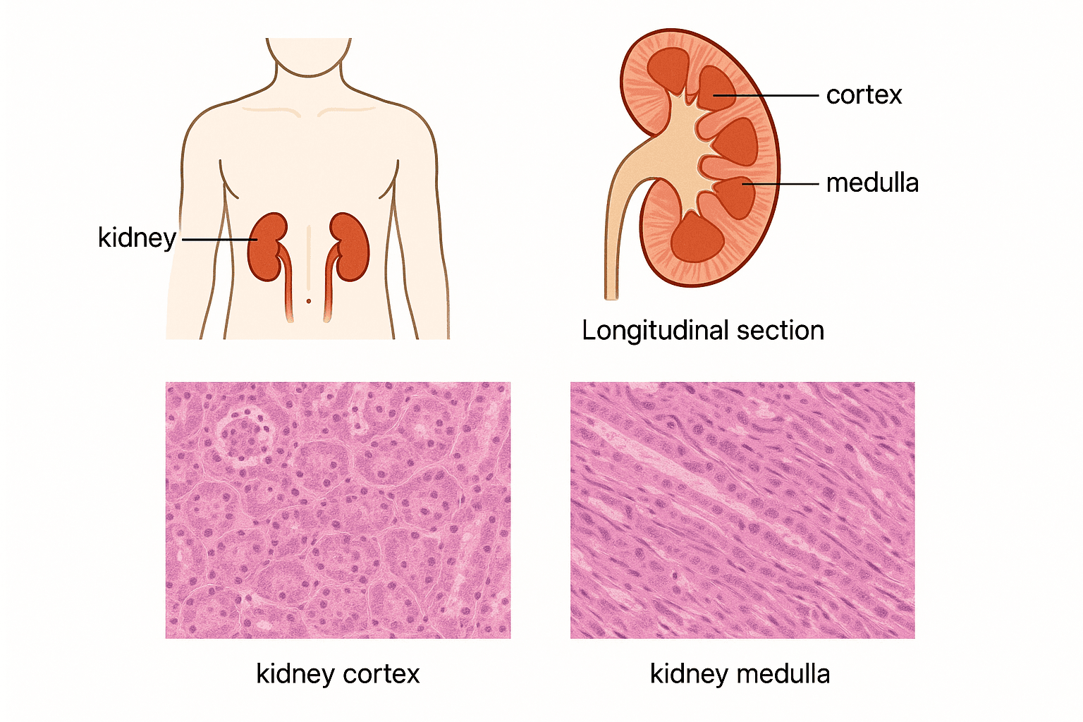
nephron — The structural and functional unit of the kidney composed of Bowman’s capsule and a tubule divided into three regions: proximal convoluted tubule, loop of Henle and distal convoluted tubule.
Each kidney contains thousands of nephrons, which are responsible for filtering blood, reabsorbing useful substances, and forming urine to regulate body fluid composition. Like a miniature water treatment plant, each nephron filters, purifies, and concentrates waste from the blood.
afferent arteriole — Arteriole leading to glomerular capillaries.
It has a wider diameter than the efferent arteriole, contributing to the high hydrostatic pressure within the glomerulus, which is essential for ultrafiltration. Like a wide pipe feeding water into a narrower hose, it creates pressure in the glomerulus.
efferent arteriole — Arteriole leading away from glomerular capillaries.
Its narrower diameter, compared to the afferent arteriole, helps maintain the high pressure in the glomerulus, facilitating ultrafiltration. Like a narrower pipe restricting water flow out of a system, it helps build up pressure upstream.
glomerulus — A group of capillaries within the ‘cup’ of a Bowman’s capsule in the cortex of the kidney.
The high pressure within these capillaries, due to the afferent arteriole being wider than the efferent, drives the process of ultrafiltration, forcing fluid and small solutes into Bowman's capsule. Like a high-pressure sprinkler head, the glomerulus forces water (blood plasma) through tiny holes (capillary pores) into the surrounding collection basin (Bowman's capsule).
Bowman’s capsule — The cup-shaped part of a nephron that surrounds a glomerulus and collects filtrate from the blood.
This is where ultrafiltration occurs, as blood plasma (minus large proteins and cells) is forced out of the glomerulus and into the capsule to form glomerular filtrate. Like a coffee filter and funnel, the Bowman's capsule collects the liquid (filtrate) that passes through the filter (glomerulus).
podocyte — One of the cells that makes up the lining of Bowman’s capsule surrounding the glomerular capillaries.
These cells have finger-like projections with gaps (slit pores) that form part of the filtration barrier, allowing filtrate to pass through while preventing the passage of larger molecules. Like a hand with fingers spread out, the podocyte's projections create spaces for fluid to pass through, but still act as a barrier.
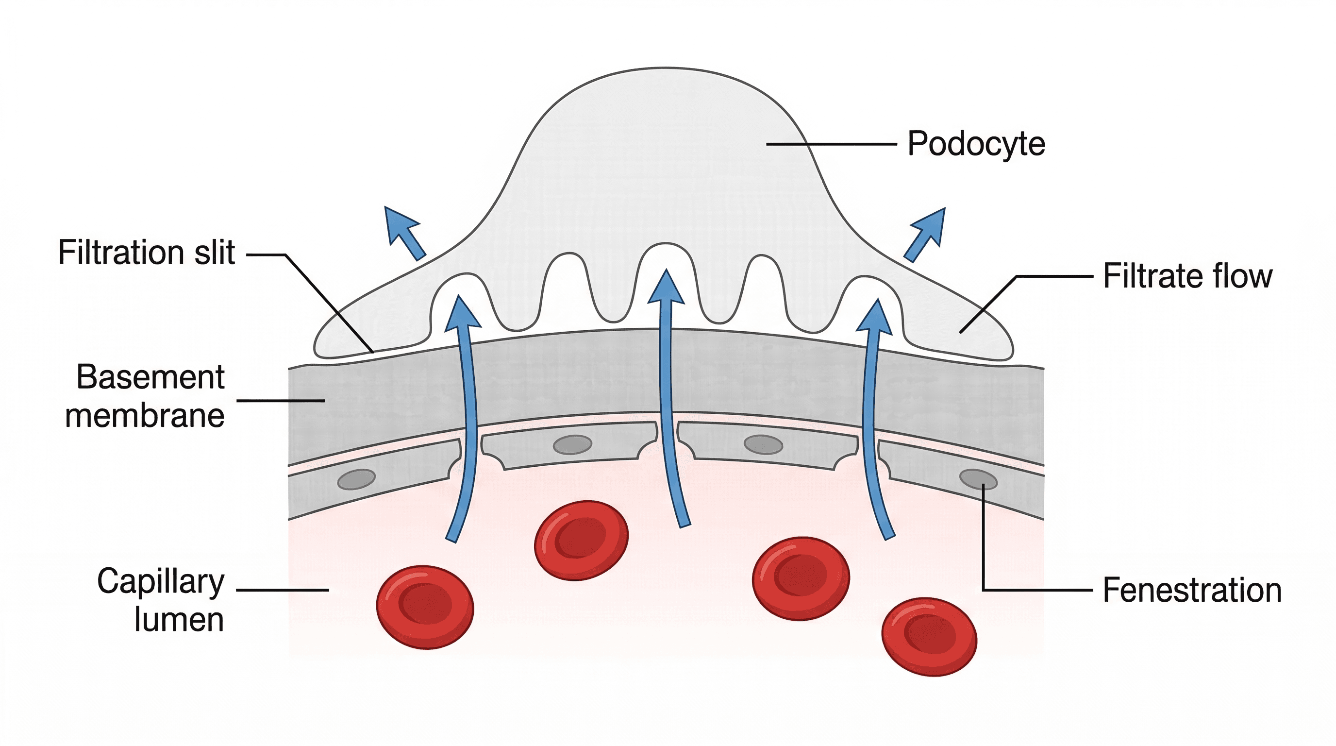
ultrafiltration — Filtration on a molecular scale separating small molecules from larger molecules, such as proteins (e.g. the filtration that occurs as blood flows through capillaries, especially those in glomeruli in the kidney).
This process occurs in the Bowman's capsule, driven by high blood pressure, forcing water and small solutes out of the blood while retaining large proteins and blood cells. Like a very fine sieve, ultrafiltration allows small particles (water, ions, glucose) to pass through but blocks larger ones (proteins, blood cells).
For ultrafiltration, always mention the high hydrostatic pressure in the glomerulus forcing small molecules from the blood into the Bowman's capsule.
selective reabsorption — Movement of certain substances from the filtrate in nephrons back into the blood.
This process ensures that useful substances like glucose, amino acids, and most water are reclaimed from the filtrate and returned to the bloodstream, preventing their loss in urine. Like a quality control check, the nephron selectively reclaims valuable items from the filtered fluid, sending them back to circulation.
proximal convoluted tubule — Part of the nephron that leads from Bowman’s capsule to the loop of Henle.
This region is responsible for the majority of selective reabsorption, taking back essential substances like glucose, amino acids, and most water from the filtrate into the blood. Like a diligent recycling plant, the proximal convoluted tubule reclaims almost all the valuable materials from the initial waste stream.
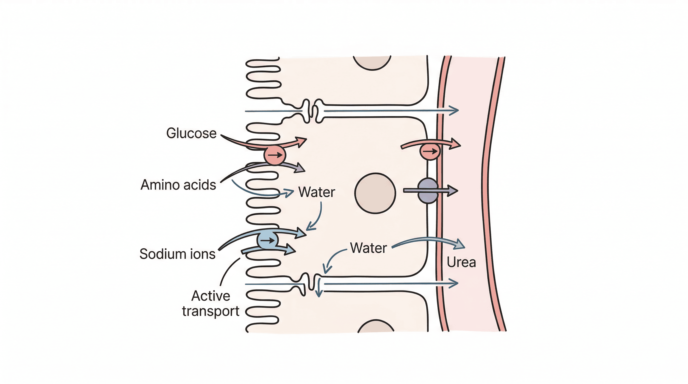
Link the structure of the proximal convoluted tubule (PCT) to its function: microvilli for large surface area and many mitochondria for active transport in selective reabsorption.
loop of Henle — The part of the nephron between the proximal and distal convoluted tubules.
Its primary function is to create a high concentration of sodium and chloride ions in the tissue fluid of the medulla, establishing a water potential gradient crucial for water reabsorption from the collecting duct. Like a counter-current heat exchanger, the loop of Henle uses opposing flows to build up a concentration gradient in the surrounding tissue.
distal convoluted tubule — Part of the nephron that leads from the loop of Henle to the collecting duct.
This region allows for fine-tuning of ion and water reabsorption, with its permeability to water being regulated by ADH, contributing to the final concentration of urine. Like the final adjustment knob on a machine, the distal convoluted tubule makes precise changes to the filtrate composition before it becomes urine.
collecting duct — Tube in the medulla of the kidney that carries urine from the distal convoluted tubules of many nephrons to the renal pelvis.
The collecting duct's permeability to water is regulated by ADH, allowing variable amounts of water to be reabsorbed by osmosis into the highly concentrated medullary tissue fluid, thus controlling urine concentration. Like a water tap that can be opened or closed, the collecting duct's permeability is controlled by ADH to adjust how much water is retained or lost.
Urine is formed in the nephrons through two main processes: ultrafiltration and selective reabsorption. Ultrafiltration occurs in the Bowman's capsule, where high hydrostatic pressure forces blood plasma (excluding large proteins and cells) into the capsule. The resulting filtrate then undergoes selective reabsorption in the proximal convoluted tubule, loop of Henle, distal convoluted tubule, and collecting duct, where useful substances are returned to the blood, and waste products are concentrated into urine.
osmoregulation — The control of the water potential of blood and tissue fluid by controlling the water content and/or the concentration of ions, particularly sodium ions.
This homeostatic process maintains the appropriate water balance in the body, preventing cells from swelling or shrinking due to osmotic imbalances, which would disrupt metabolic functions. Like a gardener carefully watering plants to keep the soil moisture just right, osmoregulation maintains the perfect water balance in the body's internal environment.
osmoreceptor — Type of receptor that detects changes in the water potential of blood.
Located in the hypothalamus, these specialised sensory neurones monitor blood water potential and initiate the release of ADH when a decrease is detected, triggering water reabsorption. Like a humidity sensor, an osmoreceptor detects changes in the 'wetness' (water potential) of the blood and signals for adjustment.
antidiuretic hormone (ADH) — Hormone secreted from the posterior pituitary gland that increases water reabsorption in the kidneys and therefore reduces water loss in urine.
ADH acts on the collecting ducts and distal convoluted tubules, increasing their permeability to water by inserting aquaporins, allowing more water to be reabsorbed into the blood when the body is dehydrated. Like a 'water-saving' signal, ADH tells the kidneys to open more water channels, allowing the body to conserve water.
Students often think ADH directly causes water to move, but actually, it increases the *permeability* of collecting ducts to water, allowing osmosis to occur down an existing water potential gradient.
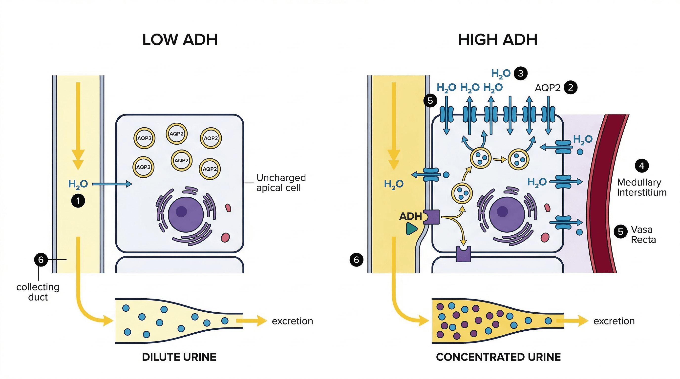
Be precise about ADH's mechanism: it binds to receptors, causing aquaporins to be inserted into the cell surface membranes of the collecting duct and DCT.
The kidneys control blood water potential through osmoregulation, a process coordinated by osmoreceptors in the hypothalamus and the hormone ADH. When blood water potential decreases, osmoreceptors stimulate the posterior pituitary gland to release ADH. ADH increases the permeability of the distal convoluted tubule and collecting duct to water, leading to increased water reabsorption and the production of more concentrated urine.
islet of Langerhans — A group of cells in the pancreas which secrete glucagon and insulin.
These endocrine clusters within the pancreas contain alpha cells (secreting glucagon) and beta cells (secreting insulin), which are crucial for the homeostatic control of blood glucose concentration. Like tiny islands of specialized factories within a larger organ, the islets produce the specific hormones needed to regulate blood sugar.
insulin — A small peptide hormone secreted by the β cells in the islets of Langerhans in the pancreas to bring about a decrease in the concentration of glucose in the blood.
Insulin stimulates liver, muscle, and adipose tissue cells to increase glucose uptake from the blood, convert glucose to glycogen (glycogenesis), and increase glucose use in respiration, thereby lowering blood glucose. Like a 'key' that unlocks cells to let glucose in, insulin helps remove excess glucose from the bloodstream.
glycogenesis — Synthesis of glycogen by addition of glucose monomers.
This process is stimulated by insulin when blood glucose levels are high, converting excess glucose into glycogen for storage primarily in the liver and muscle cells. It is a key mechanism for lowering blood glucose concentration.
glucagon — A small peptide hormone secreted by the α cells in the islets of Langerhans in the pancreas to bring about an increase in the concentration of glucose in the blood.
Glucagon primarily acts on liver cells, stimulating glycogenolysis (breakdown of glycogen) and gluconeogenesis (formation of new glucose) to release glucose into the bloodstream when blood glucose levels are low. Like an 'emergency release' button for stored energy, glucagon signals the liver to release glucose when blood sugar drops.
glycogenolysis — The breakdown of glycogen by removal of glucose monomers.
This process is stimulated by glucagon when blood glucose levels are low, releasing stored glucose from glycogen in the liver into the bloodstream. It is a crucial mechanism for increasing blood glucose concentration.
gluconeogenesis — The formation of glucose in the liver from non-carbohydrate sources such as amino acids, pyruvate, lactate, fatty acids and glycerol.
This process is stimulated by glucagon when blood glucose levels are low and glycogen stores are depleted, providing an alternative source of glucose to maintain blood sugar. It is vital for long-term glucose regulation.
adenylyl cyclase — Enzyme that catalyses formation of the second messenger cyclic AMP.
Adenylyl cyclase is a key enzyme in cell signalling pathways, particularly in the response to hormones like glucagon. Its activation leads to the production of cAMP, which then amplifies the hormonal signal within the cell.
cyclic AMP (c-AMP) — A second messenger in cell-signalling pathways.
Cyclic AMP acts as an intracellular signal, relaying and amplifying the message from a hormone (first messenger) that binds to a receptor on the cell surface. It triggers a cascade of events inside the cell, leading to the final physiological response.
protein kinase A — Enzyme that is activated by c-AMP and once activated adds phosphate groups to other proteins, including phosphorylase kinase, to activate them.
Protein kinase A is a crucial enzyme in many cell signalling pathways. Its activation by cAMP initiates a phosphorylation cascade, where it modifies other proteins by adding phosphate groups, thereby altering their activity and leading to a cellular response.
phosphorylase kinase — An enzyme that is part of the enzyme cascade that acts in response to glucagon; the enzyme activates glycogen phosphorylase by adding a phosphate group.
Phosphorylase kinase plays a specific role in the glucagon signalling pathway. Upon activation by protein kinase A, it phosphorylates and activates glycogen phosphorylase, which is the enzyme directly responsible for breaking down glycogen into glucose.
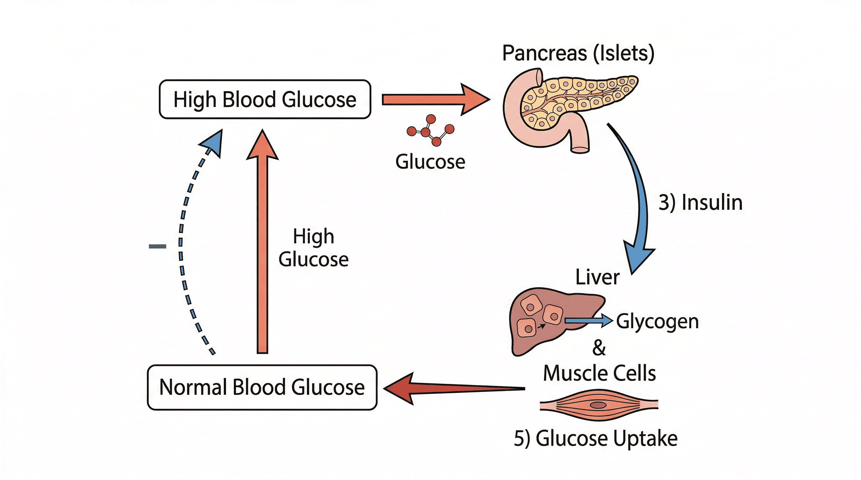
Blood glucose concentration is tightly controlled by the hormones insulin and glucagon, secreted by the islets of Langerhans in the pancreas. When blood glucose is high, beta cells release insulin, which promotes glucose uptake by liver, muscle, and adipose cells, and stimulates glycogenesis. When blood glucose is low, alpha cells release glucagon, which acts on the liver to stimulate glycogenolysis and gluconeogenesis, releasing glucose into the blood.
Students often think glucagon acts on muscle cells, but actually, muscle cells do not have glucagon receptors; its primary target for glucose release is the liver.
Clearly distinguish between insulin and glucagon effects. Use key terms: insulin promotes glycogenesis (glucose to glycogen), while glucagon promotes glycogenolysis (glycogen to glucose).
biosensor — A device that uses a biological material such as an enzyme to measure the concentration of a chemical compound.
Biosensors are used for measuring blood and urine glucose, often employing enzymes like glucose oxidase and peroxidase. They provide a rapid and accurate way to monitor physiological parameters. Their core function relies on a biological component, typically an enzyme, to detect the target substance.
Students often think biosensors are purely electronic, but actually, their core function relies on a biological component, typically an enzyme.
Test strips and biosensors are commonly used to measure glucose concentration in blood and urine. Biosensors utilise biological materials, typically enzymes like glucose oxidase and peroxidase, to detect and quantify glucose. This allows for rapid and accurate monitoring, crucial for managing conditions like diabetes.
guard cell — A kidney-shaped epidermal cell found with another, in a pair surrounding a stoma and controlling its opening or closure.
Guard cells regulate the stomatal aperture, balancing carbon dioxide entry for photosynthesis with water loss by transpiration. Their turgor changes, influenced by ion movement and water potential, dictate whether the stoma is open or closed.
electrochemical gradient — A gradient across a cell surface membrane that involves both a difference in concentrations of ions and a potential difference.
This gradient is crucial for the movement of ions, such as potassium ions, into and out of guard cells, which in turn affects their turgor and the opening or closing of stomata. It represents the combined force driving ion movement across a membrane.
abscisic acid (ABA) — An inhibitory plant growth regulator that causes closure of stomata in dry conditions.
Abscisic acid plays a vital role in plant water conservation. During water shortage, ABA signals guard cells to close stomata, reducing transpiration and preventing excessive water loss, thus helping the plant cope with drought stress.
Plants also exhibit homeostatic mechanisms, particularly in regulating stomatal aperture to balance carbon dioxide entry for photosynthesis with water loss by transpiration. Guard cells, surrounding the stomata, control their opening and closing. Stomata have daily rhythms and respond to environmental changes, such as light and water availability. During water shortage, the plant hormone abscisic acid (ABA) triggers stomatal closure to conserve water.
Use specific terminology accurately, such as 'water potential gradient', 'selective reabsorption', and 'deamination' to maximise marks.
homeostasis
The maintenance of a relatively constant internal environment for the cells within the body.
negative feedback
A process in which a change in some parameter (e.g. blood glucose concentration) brings about processes which return it towards normal.
receptor
A cell or tissue that is sensitive to a specific stimulus and communicates with a control centre by generating nerve impulses or sending a chemical messenger.
effector
A tissue or organ that carries out an action in response to a stimulus; muscles and glands are effectors.
stimulus
A change in the external or internal environment that is detected by a receptor and which may cause a response.
corrective action
A response or series of responses that return a physiological factor to the set point so maintaining a constant environment for the cells within the body.
set point
The ideal value of a physiological factor that the body controls in homeostasis.
hormone
A substance secreted by an endocrine gland that is carried in blood plasma to another part of the body where it has an effect.
positive feedback
A process in which a change in some parameter such as a physiological factor brings about processes that move its level further in the direction of the initial change.
excretion
The removal of toxic or waste products of metabolism from the body.
urea
A nitrogenous excretory product produced in the liver from the deamination of amino acids.
deamination
The breakdown of excess amino acids in the liver, by the removal of the amine group; ammonia and, eventually, urea are formed from the amine group.
nephron
The structural and functional unit of the kidney composed of Bowman’s capsule and a tubule divided into three regions: proximal convoluted tubule, loop of Henle and distal convoluted tubule.
Bowman’s capsule
The cup-shaped part of a nephron that surrounds a glomerulus and collects filtrate from the blood.
glomerulus
A group of capillaries within the ‘cup’ of a Bowman’s capsule in the cortex of the kidney.
proximal convoluted tubule
Part of the nephron that leads from Bowman’s capsule to the loop of Henle.
loop of Henle
The part of the nephron between the proximal and distal convoluted tubules.
distal convoluted tubule
Part of the nephron that leads from the loop of Henle to the collecting duct.
collecting duct
Tube in the medulla of the kidney that carries urine from the distal convoluted tubules of many nephrons to the renal pelvis.
afferent arteriole
Arteriole leading to glomerular capillaries.
efferent arteriole
Arteriole leading away from glomerular capillaries.
ultrafiltration
Filtration on a molecular scale separating small molecules from larger molecules, such as proteins (e.g. the filtration that occurs as blood flows through capillaries, especially those in glomeruli in the kidney).
selective reabsorption
Movement of certain substances from the filtrate in nephrons back into the blood.
podocyte
One of the cells that makes up the lining of Bowman’s capsule surrounding the glomerular capillaries.
osmoregulation
The control of the water potential of blood and tissue fluid by controlling the water content and/or the concentration of ions, particularly sodium ions.
osmoreceptor
Type of receptor that detects changes in the water potential of blood.
antidiuretic hormone (ADH)
Hormone secreted from the posterior pituitary gland that increases water reabsorption in the kidneys and therefore reduces water loss in urine.
islet of Langerhans
A group of cells in the pancreas which secrete glucagon and insulin.
glucagon
A small peptide hormone secreted by the α cells in the islets of Langerhans in the pancreas to bring about an increase in the concentration of glucose in the blood.
insulin
A small peptide hormone secreted by the β cells in the islets of Langerhans in the pancreas to bring about a decrease in the concentration of glucose in the blood.
glycogenesis
Synthesis of glycogen by addition of glucose monomers.
adenylyl cyclase
Enzyme that catalyses formation of the second messenger cyclic AMP.
cyclic AMP (c-AMP)
A second messenger in cell-signalling pathways.
protein kinase A
Enzyme that is activated by c-AMP and once activated adds phosphate groups to other proteins, including phosphorylase kinase, to activate them.
phosphorylase kinase
An enzyme that is part of the enzyme cascade that acts in response to glucagon; the enzyme activates glycogen phosphorylase by adding a phosphate group.
glycogenolysis
The breakdown of glycogen by removal of glucose monomers.
gluconeogenesis
The formation of glucose in the liver from non-carbohydrate sources such as amino acids, pyruvate, lactate, fatty acids and glycerol.
biosensor
A device that uses a biological material such as an enzyme to measure the concentration of a chemical compound.
guard cell
A kidney-shaped epidermal cell found with another, in a pair surrounding a stoma and controlling its opening or closure.
electrochemical gradient
A gradient across a cell surface membrane that involves both a difference in concentrations of ions and a potential difference.
abscisic acid (ABA)
An inhibitory plant growth regulator that causes closure of stomata in dry conditions.
| Command word | What examiners expect |
|---|---|
| Explain | When explaining the importance of homeostasis, link it directly to enzyme activity and metabolic efficiency, mentioning denaturation at extremes. For ADH action, explain the mechanism: binding to receptors, cAMP production, vesicle fusion, aquaporin insertion, and increased water permeability. |
| Describe | When describing negative feedback, clearly identify the stimulus, receptor, control centre, effector, and the corrective action that reverses the initial change. For deamination, specify that it occurs in the liver, involves the removal of the amine group, and produces ammonia (which is then converted to urea) and a keto acid. |
| State | State that urea is produced by deamination of excess amino acids in the liver. State the location (hypothalamus) and specific function (detecting blood water potential) of osmoreceptors. |
| Identify | When identifying effectors, name the specific organ or tissue (e.g., liver cells, muscle cells, collecting ducts) and the action they perform. Be able to label and describe the function of each part of the nephron and its associated blood vessels. |
Mistake
Thinking homeostasis means conditions are absolutely constant.
Correction
Homeostasis maintains conditions that fluctuate slightly around a set point, not absolute constancy.
Mistake
Confusing negative feedback with a 'bad' response.
Correction
Negative feedback refers to a response that reverses the initial change, maintaining stability, which is beneficial.
Mistake
Confusing excretion with egestion.
Correction
Excretion is the removal of metabolic waste products (e.g., urea), while egestion is the removal of undigested food.
Mistake
Believing ADH directly causes water to move.
Correction
ADH increases the *permeability* of collecting ducts to water, allowing osmosis to occur down an existing water potential gradient.
Mistake
Thinking glucagon acts on muscle cells.
Correction
Muscle cells do not have glucagon receptors; its primary target for glucose release is the liver.
Mistake
Assuming biosensors are purely electronic.
Correction
The core function of biosensors relies on a biological component, typically an enzyme.