Biology · Control and coordination
This chapter explores the intricate mechanisms of control and coordination in living organisms, focusing on the endocrine and nervous systems in mammals, and electrical and chemical communication in plants. It details the structure and function of neurones, the transmission of nerve impulses via action potentials and synapses, and the ultrastructure and contraction mechanism of striated muscle. Additionally, it covers rapid plant responses and the roles of plant growth regulators such as auxins and gibberellins.
endocrine system — Consists of all the endocrine glands in the body together with the hormones that they secrete.
This system is one of the body's main communication and coordination systems, working alongside the nervous system to regulate various physiological processes, often over longer durations. It is like the body's slow-acting, widespread postal service, delivering chemical messages (hormones) to many different locations.
endocrine gland — An organ that secretes hormones directly into the blood; endocrine glands are also known as ductless glands.
These glands are part of the endocrine system and release their chemical messengers (hormones) into the bloodstream, allowing them to travel to distant target cells throughout the body. This contrasts with exocrine glands, which secrete substances through ducts. Think of an endocrine gland as a radio station broadcasting a signal (hormone) to anyone with a receiver (receptor) tuned to that frequency, rather than sending a letter (exocrine) to a specific address.
When asked to describe endocrine glands, always mention 'ductless' and 'secrete hormones directly into the blood' for full marks.
Students often think endocrine glands have ducts, but actually they are ductless and secrete directly into the blood.
neurone — A nerve cell; a cell which is specialised for the conduction of nerve impulses.
Neurones are the basic functional units of the nervous system, designed to transmit electrical signals rapidly over long distances to coordinate body activities. A neurone is like a telegraph wire, specifically designed to carry electrical signals (impulses) from one point to another very quickly.
nerve impulse — (usually shortened to impulse) a wave of electrical depolarisation that is transmitted along neurones.
This is the fundamental unit of information transfer in the nervous system, involving rapid changes in the electrical potential across the neurone's cell surface membrane due to ion movement. A nerve impulse is like a ripple effect in a pond, where the disturbance (depolarisation) travels along the surface (neurone membrane) without the water itself moving far.
Students often think a nerve impulse is a flow of electrons like an electric current, but actually it's a wave of electrical depolarisation caused by ion movement.
Avoid using 'electrical current' when describing nerve impulses; instead, use 'wave of electrical depolarisation' or 'action potential' and mention ion movement.
The endocrine and nervous systems are the body's main communication and coordination systems. The endocrine system uses chemical messengers (hormones) transported by the blood, resulting in slower, widespread, and often longer-lasting responses. In contrast, the nervous system uses electrical impulses transmitted along neurones, leading to rapid, targeted, and short-lived responses.
When comparing the endocrine and nervous systems, focus on the speed and duration of response, and the mode of transport (blood vs. electrical impulses).
sensory neurone — A neurone that transmits nerve impulses from a receptor to the central nervous system (CNS).
These neurones are responsible for conveying information about internal and external stimuli from sensory receptors to the brain and spinal cord for processing. A sensory neurone is like a security camera cable, sending signals from a sensor (receptor) to the central monitoring station (CNS).
motor neurone — A neurone whose cell body is in the brain, spinal cord or a ganglion (a swelling on a nerve), and that transmits nerve impulses to an effector such as a muscle or gland.
Motor neurones are crucial for initiating responses by carrying commands from the CNS to muscles or glands, causing them to contract or secrete. A motor neurone is like a control wire from a central command center (CNS) to a robot's arm (effector), telling it to move.
intermediate neurone — A neurone that transmits nerve impulses between other neurones, typically within the central nervous system (CNS).
Intermediate neurones, also known as relay neurones, connect sensory and motor neurones, allowing for complex processing and integration of signals within the CNS. They are essential for reflex arcs and higher-order brain functions.
Remember that sensory neurones carry information 'to' the CNS, while motor neurones carry information 'from' the CNS.
Students often think all neurones are the same, but actually there are different types (sensory, motor, intermediate) with distinct structures and functions.
myelin — Insulating material that surrounds the axons of many neurones; myelin is made of layers of cell surface membranes formed by Schwann cells so that they are very rich in phospholipids and therefore impermeable to water and ions in tissue fluid.
Myelin acts as an electrical insulator, significantly increasing the speed of nerve impulse conduction by allowing action potentials to 'jump' between nodes of Ranvier. Myelin is like the plastic insulation around an electrical wire, preventing signal leakage and allowing the electrical impulse to travel much faster.
node of Ranvier — A very short gap between Schwann cells where myelinated axons are not covered in myelin so are exposed to tissue fluid.
These gaps are critical for saltatory conduction, as action potentials are regenerated only at these points, allowing the impulse to 'jump' from node to node. Nodes of Ranvier are like stepping stones across a river; the impulse jumps from one stone to the next, rather than having to wade through the entire river.
Explain that myelin is rich in phospholipids, making it impermeable to ions, and that it enables saltatory conduction for faster impulse transmission.
Students often think myelin is a continuous sheath, but actually it has small gaps called nodes of Ranvier where depolarisation occurs.
The resting potential is the electrical potential difference across the cell surface membrane of a neurone when it is not transmitting an action potential, typically around -70 mV inside. This negative charge is established and maintained by the sodium-potassium pump, which actively transports three sodium ions out for every two potassium ions in, along with the differential permeability of the membrane to ions and the presence of large organic anions inside the cell. This state prepares the neurone to fire an action potential.
potential difference — The difference in electrical potential between two points; in the nervous system, between the inside and the outside of a cell surface membrane such as the membrane that encloses an axon.
This electrical gradient is crucial for nerve impulse transmission, as changes in potential difference across the membrane drive the opening and closing of voltage-gated ion channels. Potential difference is like the height difference between two points on a hill; it creates a 'drive' for things (like ions) to move from higher to lower potential.
resting potential — The difference in electrical potential that is maintained across the cell surface membrane of a neurone when it is not transmitting an action potential; it is normally about –70 mV inside and is partly maintained by sodium–potassium pumps.
This negative charge inside the neurone is established and maintained by the sodium-potassium pump, differential ion permeability, and the presence of large organic anions, preparing the neurone to fire an action potential. The resting potential is like a loaded spring, storing potential energy (electrical charge) that can be released quickly when triggered (by a stimulus).
Students often think the resting potential is solely due to the sodium-potassium pump, but actually the differential permeability of the membrane to ions and large intracellular anions also contribute significantly.
When explaining resting potential, mention the roles of the sodium-potassium pump, the impermeability of the membrane to sodium ions, and the presence of organic anions.
A nerve impulse is transmitted as an action potential, a rapid, brief change in the potential difference across the neurone membrane. This 'all-or-none' event is triggered when a stimulus causes the membrane potential to reach a critical threshold potential. Once initiated, the action potential propagates along the neurone, involving sequential depolarisation and repolarisation.
action potential — A brief change in the potential difference from –70 mV to +30 mV across the cell surface membranes of neurones and muscle cells caused by the inward movement of sodium ions.
This is the electrical signal transmitted along a neurone, involving a rapid depolarisation followed by repolarisation, driven by the sequential opening and closing of voltage-gated ion channels. An action potential is like flipping a light switch on and off very quickly; the voltage rapidly changes from negative to positive and back again.
voltage-gated channel protein — A channel protein through a cell membrane that opens or closes in response to changes in electrical potential across the membrane.
These proteins are fundamental to action potential generation and propagation, as their opening and closing in response to depolarisation allows the rapid influx and efflux of ions. A voltage-gated channel protein is like an automatic door that only opens when a specific electrical 'key' (voltage change) is detected.
depolarisation — The reversal of the resting potential across the cell surface membrane of a neurone or muscle cell, so that the inside becomes positively charged compared with the outside.
This is the initial phase of an action potential, caused by the rapid influx of sodium ions through voltage-gated channels, making the inside of the membrane less negative and then positive. Depolarisation is like a sudden surge of water into a dry well, quickly filling it up and even overflowing, changing its 'potential' from empty to full.
threshold potential — The critical potential difference across the cell surface membrane of a sensory receptor or neurone which must be reached before an action potential is initiated.
This 'all-or-none' principle means that if the stimulus is too weak to reach this threshold, no action potential will be generated, ensuring that only significant stimuli trigger responses. The threshold potential is like the minimum pressure needed to push a button; if you don't push hard enough, nothing happens, but once you reach that pressure, the button activates fully.
repolarisation — Returning the potential difference across the cell surface membrane of a neurone or muscle cell to normal following the depolarisation of an action potential.
This phase involves the efflux of potassium ions through voltage-gated channels, restoring the negative charge inside the membrane and preparing the neurone for another action potential. Repolarisation is like draining the overflowing well back to its original dry state, restoring its 'potential' to be filled again.
refractory period — A period of time during which a neurone is recovering from an action potential, and during which another action potential cannot be generated.
This period ensures that action potentials are discrete events, travel in one direction only, and limits the maximum frequency at which impulses can be transmitted. The refractory period is like the cool-down time for a camera flash; you can't take another picture immediately after one flash, you have to wait for it to recharge.
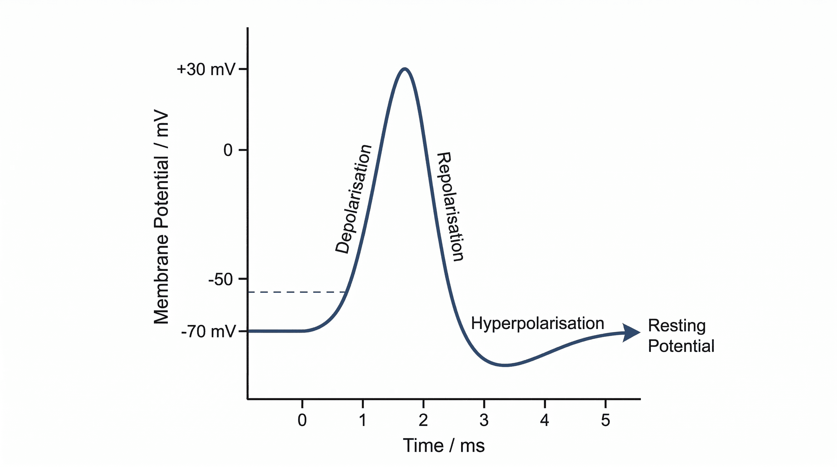
Clearly state the voltage changes (e.g., -70mV to +30mV) and the primary ion responsible for depolarisation (sodium ions) when describing an action potential.
Students often think action potentials vary in size with stimulus strength, but actually they are 'all-or-none' events with a constant amplitude; only their frequency changes.
all-or-none law — Neurones and muscle cells only transmit impulses if the initial stimulus is sufficient to increase the membrane potential above a threshold potential.
This principle dictates that once the threshold is reached, an action potential of a fixed amplitude is generated, regardless of further increases in stimulus strength; information about stimulus intensity is encoded by frequency. The all-or-none law is like flushing a toilet; once you push the handle past a certain point, the flush happens completely, regardless of how much harder you push.
Emphasise the 'all-or-none' nature of action potentials in relation to the threshold potential; a sub-threshold stimulus produces no action potential.
The speed of nerve impulse conduction is significantly increased in myelinated neurones through a process called saltatory conduction. Myelin acts as an electrical insulator, preventing ion flow across the membrane except at the nodes of Ranvier. This allows the action potential to 'jump' from one node to the next, rather than propagating continuously along the entire axon.
saltatory conduction — Movement of an action potential along a myelinated axon, in which the action potential ‘jumps’ from one node of Ranvier to the next.
This mechanism significantly increases the speed of impulse transmission in myelinated neurones compared to unmyelinated ones, as depolarisation only occurs at the nodes. Saltatory conduction is like skipping steps on a staircase instead of taking each one; it's a much faster way to get from top to bottom.
Explain that myelin insulates the axon, preventing ion flow, and that voltage-gated channels are concentrated at the nodes, allowing the 'jumping' effect.
Students often think saltatory conduction means the impulse literally jumps through the air, but actually it means the action potential is regenerated only at the nodes of Ranvier.
chemoreceptor — A receptor cell that responds to chemical stimuli; chemoreceptors are found in taste buds on the tongue, in the nose and in blood vessels where they detect changes in oxygen and carbon dioxide concentrations.
These specialised cells convert chemical signals into electrical impulses, enabling senses like taste and smell, and playing vital roles in homeostatic regulation by monitoring blood chemistry. A chemoreceptor is like a chemical sensor that detects specific molecules in its environment and then sends an alarm signal (electrical impulse) when it finds them.
receptor potential — A change in the normal resting potential across the membrane of a receptor cell, caused by a stimulus.
This is a graded potential, meaning its magnitude depends on the strength of the stimulus; if it reaches a threshold, it can trigger an action potential in an associated sensory neurone. A receptor potential is like the initial push on a swing; a small push causes a small swing, but a strong push can make it swing high enough to trigger a larger event (like an action potential).
Students often think receptor potentials are always action potentials, but actually receptor potentials are graded and only trigger action potentials if they reach a threshold.
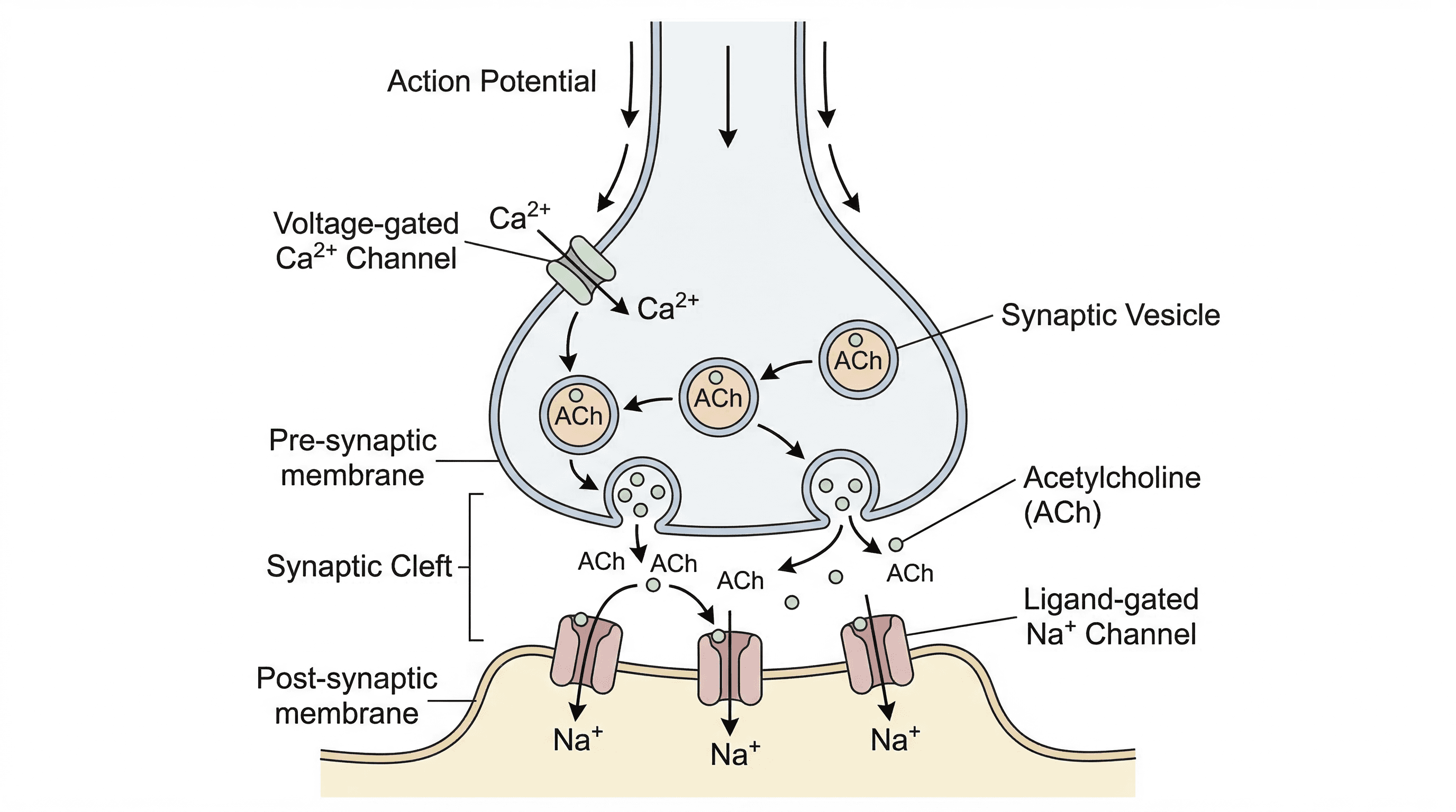
Synapses are crucial junctions where nerve impulses are transmitted from one neurone to another, or from a neurone to an effector. Unlike direct electrical connections, synapses involve a small gap called the synaptic cleft, across which chemical messengers called neurotransmitters diffuse. This chemical transmission allows for signal integration, modulation, and ensures unidirectional flow of information.
synapse — A point at which two neurones meet but do not touch; the synapse is made up of the end of the presynaptic neurone, the synaptic cleft and the end of the postsynaptic neurone.
Synapses are crucial for information processing in the nervous system, allowing for one-way transmission, integration of multiple signals, and modulation of nerve impulses. A synapse is like a junction box in an electrical circuit, where signals can be routed, amplified, or inhibited before being passed on.
synaptic cleft — A very small gap between two neurones at a synapse; nerve impulses are transmitted across synaptic clefts by neurotransmitters.
This physical separation prevents direct electrical transmission, necessitating chemical transmission via neurotransmitters, which allows for signal integration and modulation. The synaptic cleft is like a small river separating two banks; the message (neurotransmitter) has to be ferried across to reach the other side.
Students often think neurones touch at a synapse, but actually there is a distinct gap, the synaptic cleft, which neurotransmitters must cross.
neurotransmitter — A chemical released at synapses to transmit impulses between neurones or between a motor neurone and a muscle fibre.
These chemical messengers diffuse across the synaptic cleft and bind to receptors on the postsynaptic membrane, causing a change in its potential and potentially generating a new action potential. A neurotransmitter is like a chemical key that unlocks a specific door (receptor) on the next cell, allowing a message to pass through.
presynaptic neurone — A neurone ending at a synapse from which neurotransmitter is released when an action potential arrives.
This neurone transmits the signal towards the synapse, releasing neurotransmitters into the synaptic cleft upon the arrival of an action potential. The presynaptic neurone is like the sender of a text message, initiating the communication by releasing the message (neurotransmitter).
postsynaptic neurone — The neurone on the opposite side of a synapse to the neurone in which the action potential arrives.
This neurone receives the neurotransmitter signal, which can cause depolarisation and potentially trigger its own action potential, propagating the impulse. The postsynaptic neurone is like the receiver of a text message, interpreting the message (neurotransmitter binding) and deciding how to respond.
voltage-gated calcium ion channel protein — A channel protein in presynaptic membranes that responds to depolarisation by opening to allow diffusion of calcium ions down their electrochemical gradient.
The influx of calcium ions through these channels into the presynaptic terminal is the critical trigger for the exocytosis of neurotransmitter vesicles. This channel is like a gate that only opens when an electrical signal arrives, letting in calcium ions which then act as a signal to release neurotransmitters.
receptor protein — A protein on a postsynaptic membrane that is a ligand-gated channel protein opening in response to binding of a neurotransmitter.
These proteins specifically bind neurotransmitters, leading to a conformational change that opens an ion channel, allowing ions to flow across the postsynaptic membrane and alter its potential. A receptor protein is like a specific lock on the postsynaptic membrane that only opens when the correct key (neurotransmitter) fits into it.
acetylcholine (ACh) — A type of neurotransmitter released by cholinergic synapses.
ACh is a common neurotransmitter involved in muscle contraction (at neuromuscular junctions), learning, and memory, and its action is rapidly terminated by acetylcholinesterase. ACh is like a specific key for muscle contraction; when it binds to the lock (receptor), the muscle is told to contract.
cholinergic synapse — A synapse at which the transmitter substance is ACh.
These synapses are prevalent in the peripheral nervous system, particularly at neuromuscular junctions, and are critical for voluntary muscle control. A cholinergic synapse is like a specific type of communication channel that only uses ACh as its language.
acetylcholinesterase — An enzyme in the synaptic cleft and on the postsynaptic membrane that hydrolyses ACh to acetate and choline.
This enzyme rapidly breaks down acetylcholine, ensuring that the neurotransmitter's effect is brief and allowing the postsynaptic membrane to repolarise and be ready for subsequent signals. Its action is crucial for precise control of muscle contraction and nerve impulse transmission.
noradrenaline — A type of neurotransmitter, which is also released by cells in the adrenal glands as a hormone.
Noradrenaline acts as both a neurotransmitter in the nervous system and a hormone in the endocrine system, playing roles in the 'fight or flight' response, alertness, and mood. Noradrenaline is like a versatile messenger that can be delivered quickly and locally (neurotransmitter) or broadcast widely through the bloodstream (hormone).
Always mention acetylcholinesterase when discussing ACh, as its role in breaking down ACh is crucial for proper synaptic function.
Muscle contraction is a complex process initiated by nerve impulses from motor neurones. Striated muscle, found in skeletal muscles, exhibits a characteristic banded appearance due to the organised arrangement of contractile proteins. The contraction itself occurs via the sliding filament model, where thick and thin filaments slide past each other, shortening the sarcomere.
neuromuscular junction — A synapse between a motor neurone and a muscle.
This specialised synapse is where the motor neurone transmits its electrical signal to the muscle fibre, initiating muscle contraction. It is a type of cholinergic synapse, using acetylcholine as its neurotransmitter.
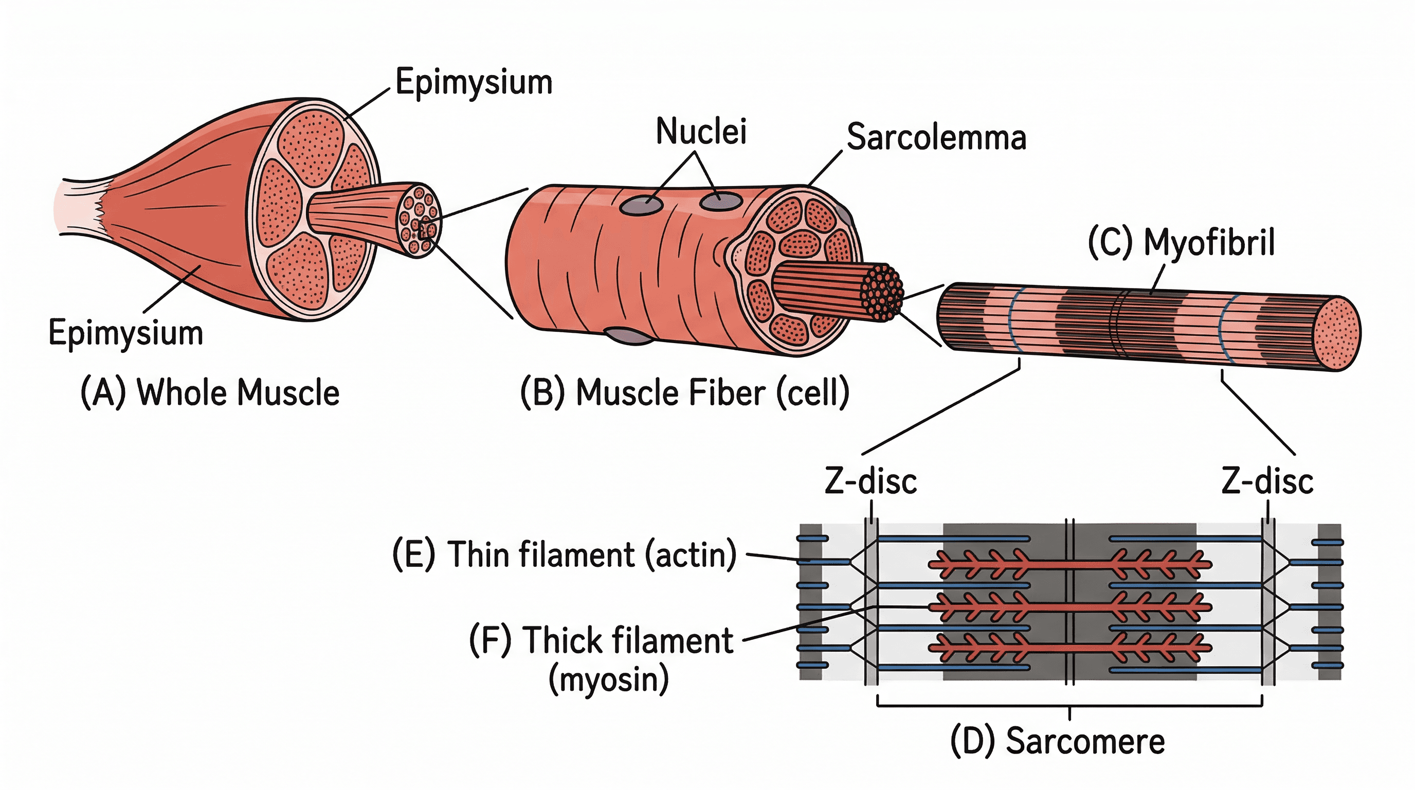
striated muscle — Type of muscle tissue in skeletal muscles; the muscle fibres have regular striations that can be seen under the light microscope.
Striated muscle is responsible for voluntary movements and is characterised by its organised structure, which gives it a striped appearance under a microscope. This organisation is key to its efficient contractile function.
sarcolemma — The cell surface membrane of a muscle fibre.
The sarcolemma is the plasma membrane of a muscle cell, which plays a crucial role in transmitting the electrical impulse from the neuromuscular junction throughout the muscle fibre via T-tubules, initiating contraction.
sarcoplasm — The cytoplasm of muscle cells.
The sarcoplasm is the specialised cytoplasm of muscle fibres, containing numerous mitochondria for ATP production, glycogen stores, and a high concentration of calcium ions, which are essential for muscle contraction.
sarcoplasmic reticulum (SR) — The endoplasmic reticulum of a muscle fibre.
The sarcoplasmic reticulum is a specialised network of membranes within muscle cells that stores and releases calcium ions, which are critical for triggering muscle contraction. It plays a central role in regulating the availability of calcium for the contractile proteins.
transverse system tubule (or T-system tubule or T-tubule) — Infolding of the sarcolemma that go deep into a muscle fibre and conducts impulses to the SR.
T-tubules are invaginations of the sarcolemma that extend deep into the muscle fibre, allowing the action potential to rapidly reach all parts of the muscle cell, including the sarcoplasmic reticulum, ensuring coordinated contraction.
myofibril — One of many cylindrical bundles of thick (myosin) and thin (actin) filaments inside a muscle fibre.
Myofibrils are the contractile units within muscle fibres, composed of repeating units called sarcomeres. Their organised arrangement of actin and myosin filaments is responsible for muscle contraction.
myosin — The protein that makes up the thick filaments in striated muscle; the globular heads of each molecule break down ATP (they act as an ATP-ase).
Myosin is a motor protein that forms the thick filaments in muscle. Its globular heads bind to actin and use ATP hydrolysis to generate the force for muscle contraction, pulling the thin filaments.
actin — The protein that makes up the thin filaments in striated muscle.
Actin is a globular protein that polymerises to form the thin filaments in muscle. It provides the binding sites for myosin heads during muscle contraction.
sarcomere — The part of a myofibril between two Z discs.
The sarcomere is the fundamental contractile unit of striated muscle, extending from one Z-disc to the next. It contains the organised arrangement of actin and myosin filaments that slide past each other during contraction.
tropomyosin — A fibrous protein that is part of the thin filaments in myofibrils in striated muscle; tropomyosin blocks the attachment site on the thin filament for myosin heads so preventing the formation of cross-bridges.
Tropomyosin is a regulatory protein that, in resting muscle, covers the myosin-binding sites on actin, preventing contraction. Its movement, triggered by calcium, exposes these sites, allowing myosin to bind.
troponin — A calcium-binding protein that is part of the thin filaments in myofibrils in striated muscle.
Troponin is a complex of three proteins that binds to calcium ions. This binding causes a conformational change in troponin, which in turn moves tropomyosin away from the myosin-binding sites on actin, initiating muscle contraction.
sliding filament model — The mechanism of muscle contraction; within each sarcomere the movement of thin filaments closer together by the action of myosin heads in the thick filaments shortens the overall length of each muscle fibre.
This model explains how muscle contraction occurs: myosin heads bind to actin filaments, form cross-bridges, and then pivot, pulling the actin filaments towards the centre of the sarcomere. This process shortens the sarcomere without the individual filaments themselves changing length.
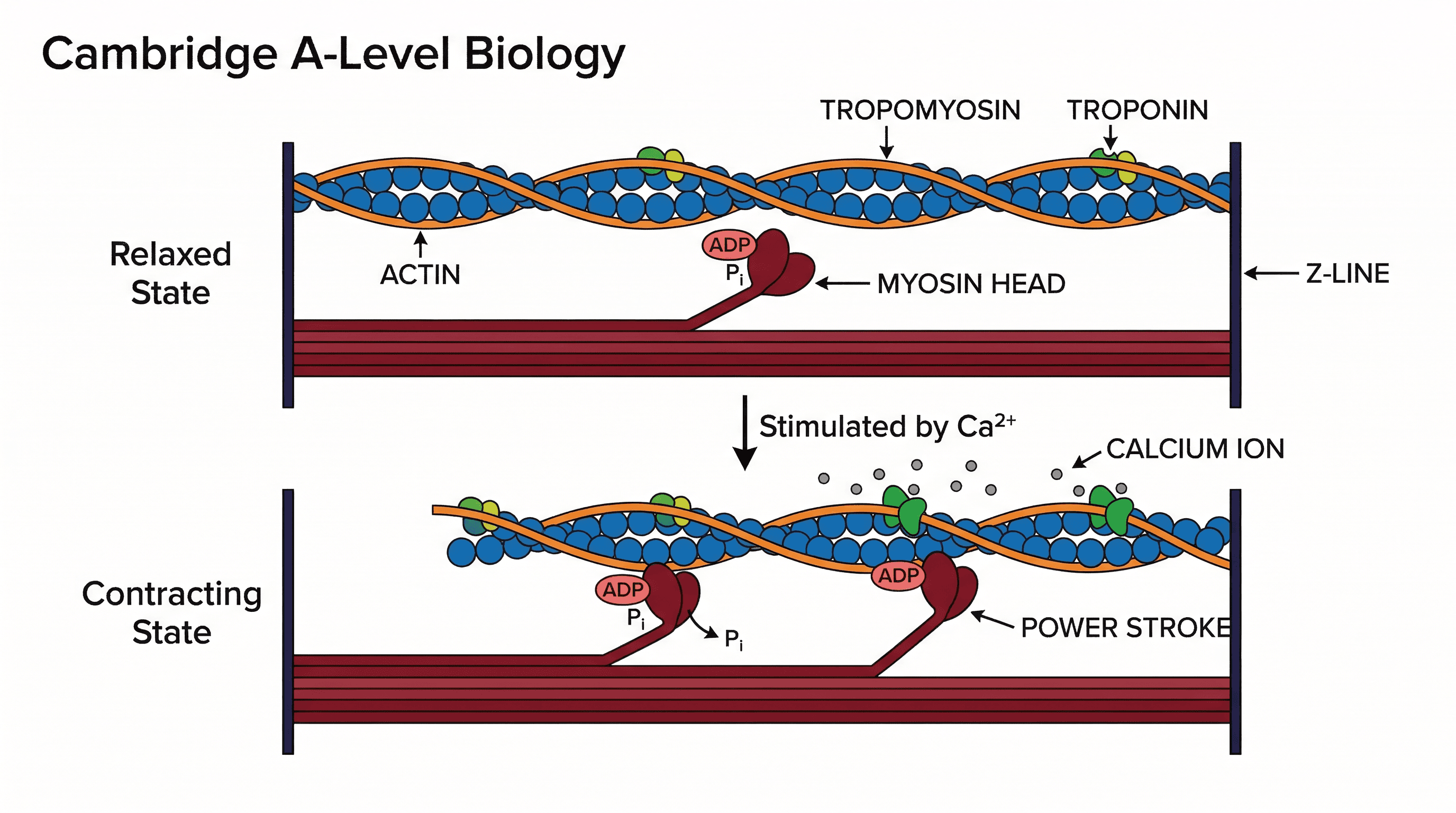
Students often think the actin and myosin filaments shorten during contraction, but actually they maintain their length and slide past each other.
For muscle contraction, be able to label a sarcomere diagram (Z-line, I-band, A-band, H-zone) and clearly state which bands shorten (I-band, H-zone) and which stay the same length (A-band).
Plants also exhibit control and coordination, though their mechanisms differ from animals. They respond to stimuli using both electrical and chemical communication. Rapid responses, such as the Venus fly trap closing, involve electrical signals. Chemical communication relies on plant growth regulators, often called plant hormones, which influence growth and development.
Students often think plant hormones are produced in specialised glands like animal hormones, but actually they are produced in various tissues throughout the plant.
plant growth regulator — (plant hormone) any chemical produced in plants that influences their growth and development (e.g. auxins, gibberellins, cytokinins and ABA).
These chemical messengers are produced in various plant tissues and regulate a wide range of physiological processes, including cell division, elongation, differentiation, and responses to environmental cues. They are crucial for coordinating plant growth and development.
auxin — A plant growth regulator (plant hormone) that stimulates cell elongation.
Auxins are primarily involved in cell elongation, particularly in shoots, and play roles in phototropism, gravitropism, and apical dominance. They promote the loosening of cell walls, allowing cells to expand.
expansins — Proteins in the cell walls of plants that loosen the attachment of microfibrils of cellulose during elongation growth.
Expansins are cell wall proteins that facilitate cell expansion by loosening the cellulose microfibril network, allowing the cell wall to stretch under turgor pressure. Auxins promote the activity of expansins, contributing to cell elongation.
gibberellin — A plant growth regulator (plant hormone) that stimulates seed germination and regulates plant height (stem growth); a lack of gibberellin causes dwarfness.
Gibberellins are a class of plant hormones involved in various developmental processes, including stem elongation, fruit development, and particularly, the breaking of seed dormancy and promotion of germination. They are crucial for normal plant growth.
endosperm — A tissue in some seeds, such as barley, that is a store of starch and other nutrients.
The endosperm serves as a primary food reserve for the developing embryo in many seeds, providing energy and building blocks until the seedling can photosynthesise independently. In barley, it is rich in starch.
aleurone layer — A layer of tissue around the endosperm that synthesises amylase during germination.
The aleurone layer is a specialised tissue in cereal grains that responds to gibberellins during germination by synthesising and secreting alpha-amylase, an enzyme that breaks down the starch in the endosperm into usable sugars for the embryo.
During barley seed germination, the embryo releases gibberellins. These gibberellins diffuse to the aleurone layer, stimulating it to synthesise and secrete alpha-amylase. Alpha-amylase then hydrolyses the starch stored in the endosperm into maltose, which is further broken down into glucose to provide energy for the growing embryo.
Always link structure to function. For example, the myelin sheath acts as an electrical insulator, and the many mitochondria in a presynaptic knob provide ATP for neurotransmitter synthesis.
Use precise terminology. Distinguish between 'neurone' (a cell) and 'nerve' (a bundle of axons). Use terms like 'presynaptic terminal', 'synaptic cleft', and 'postsynaptic membrane' correctly.
Be prepared for comparison questions. Create a table comparing nervous vs. endocrine control based on speed, duration, transmission method (neurones vs. blood), and nature of the message (electrical vs. chemical).
endocrine gland
An organ that secretes hormones directly into the blood; endocrine glands are also known as ductless glands.
endocrine system
Consists of all the endocrine glands in the body together with the hormones that they secrete.
nerve impulse
(usually shortened to impulse) a wave of electrical depolarisation that is transmitted along neurones.
neurone
A nerve cell; a cell which is specialised for the conduction of nerve impulses.
sensory neurone
A neurone that transmits nerve impulses from a receptor to the central nervous system (CNS).
motor neurone
A neurone whose cell body is in the brain, spinal cord or a ganglion (a swelling on a nerve), and that transmits nerve impulses to an effector such as a muscle or gland.
intermediate neurone
A neurone that transmits nerve impulses between other neurones, typically within the central nervous system (CNS).
myelin
Insulating material that surrounds the axons of many neurones; myelin is made of layers of cell surface membranes formed by Schwann cells so that they are very rich in phospholipids and therefore impermeable to water and ions in tissue fluid.
node of Ranvier
A very short gap between Schwann cells where myelinated axons are not covered in myelin so are exposed to tissue fluid.
action potential
A brief change in the potential difference from –70 mV to +30 mV across the cell surface membranes of neurones and muscle cells caused by the inward movement of sodium ions.
potential difference
The difference in electrical potential between two points; in the nervous system, between the inside and the outside of a cell surface membrane such as the membrane that encloses an axon.
resting potential
The difference in electrical potential that is maintained across the cell surface membrane of a neurone when it is not transmitting an action potential; it is normally about –70 mV inside and is partly maintained by sodium–potassium pumps.
voltage-gated channel protein
A channel protein through a cell membrane that opens or closes in response to changes in electrical potential across the membrane.
depolarisation
The reversal of the resting potential across the cell surface membrane of a neurone or muscle cell, so that the inside becomes positively charged compared with the outside.
threshold potential
The critical potential difference across the cell surface membrane of a sensory receptor or neurone which must be reached before an action potential is initiated.
repolarisation
Returning the potential difference across the cell surface membrane of a neurone or muscle cell to normal following the depolarisation of an action potential.
refractory period
A period of time during which a neurone is recovering from an action potential, and during which another action potential cannot be generated.
saltatory conduction
Movement of an action potential along a myelinated axon, in which the action potential ‘jumps’ from one node of Ranvier to the next.
chemoreceptor
A receptor cell that responds to chemical stimuli; chemoreceptors are found in taste buds on the tongue, in the nose and in blood vessels where they detect changes in oxygen and carbon dioxide concentrations.
receptor potential
A change in the normal resting potential across the membrane of a receptor cell, caused by a stimulus.
all-or-none law
Neurones and muscle cells only transmit impulses if the initial stimulus is sufficient to increase the membrane potential above a threshold potential.
synaptic cleft
A very small gap between two neurones at a synapse; nerve impulses are transmitted across synaptic clefts by neurotransmitters.
synapse
A point at which two neurones meet but do not touch; the synapse is made up of the end of the presynaptic neurone, the synaptic cleft and the end of the postsynaptic neurone.
neurotransmitter
A chemical released at synapses to transmit impulses between neurones or between a motor neurone and a muscle fibre.
presynaptic neurone
A neurone ending at a synapse from which neurotransmitter is released when an action potential arrives.
postsynaptic neurone
The neurone on the opposite side of a synapse to the neurone in which the action potential arrives.
noradrenaline
A type of neurotransmitter, which is also released by cells in the adrenal glands as a hormone.
acetylcholine (ACh)
A type of neurotransmitter released by cholinergic synapses.
cholinergic synapse
A synapse at which the transmitter substance is ACh.
voltage-gated calcium ion channel protein
A channel protein in presynaptic membranes that responds to depolarisation by opening to allow diffusion of calcium ions down their electrochemical gradient.
receptor protein
A protein on a postsynaptic membrane that is a ligand-gated channel protein opening in response to binding of a neurotransmitter.
acetylcholinesterase
An enzyme in the synaptic cleft and on the postsynaptic membrane that hydrolyses ACh to acetate and choline.
neuromuscular junction
A synapse between a motor neurone and a muscle.
striated muscle
Type of muscle tissue in skeletal muscles; the muscle fibres have regular striations that can be seen under the light microscope.
sarcolemma
The cell surface membrane of a muscle fibre.
sarcoplasm
The cytoplasm of muscle cells.
sarcoplasmic reticulum (SR)
The endoplasmic reticulum of a muscle fibre.
transverse system tubule (or T-system tubule or T-tubule)
Infolding of the sarcolemma that go deep into a muscle fibre and conducts impulses to the SR.
myofibril
One of many cylindrical bundles of thick (myosin) and thin (actin) filaments inside a muscle fibre.
myosin
The protein that makes up the thick filaments in striated muscle; the globular heads of each molecule break down ATP (they act as an ATP-ase).
actin
The protein that makes up the thin filaments in striated muscle.
sarcomere
The part of a myofibril between two Z discs.
tropomyosin
A fibrous protein that is part of the thin filaments in myofibrils in striated muscle; tropomyosin blocks the attachment site on the thin filament for myosin heads so preventing the formation of cross-bridges.
troponin
A calcium-binding protein that is part of the thin filaments in myofibrils in striated muscle.
sliding filament model
The mechanism of muscle contraction; within each sarcomere the movement of thin filaments closer together by the action of myosin heads in the thick filaments shortens the overall length of each muscle fibre.
plant growth regulator
(plant hormone) any chemical produced in plants that influences their growth and development (e.g. auxins, gibberellins, cytokinins and ABA).
auxin
A plant growth regulator (plant hormone) that stimulates cell elongation.
gibberellin
A plant growth regulator (plant hormone) that stimulates seed germination and regulates plant height (stem growth); a lack of gibberellin causes dwarfness.
expansins
Proteins in the cell walls of plants that loosen the attachment of microfibrils of cellulose during elongation growth.
endosperm
A tissue in some seeds, such as barley, that is a store of starch and other nutrients.
aleurone layer
A layer of tissue around the endosperm that synthesises amylase during germination.
| Command word | What examiners expect |
|---|---|
| Describe | Provide a detailed account of the features or process, e.g., 'Describe the features of the endocrine system' requires mentioning ductless glands, hormone secretion into blood, and widespread action. |
| Explain | Give reasons for a phenomenon or process, e.g., 'Explain the transmission of nerve impulses' requires detailing ion movements, voltage changes, and the role of channels. |
| Compare | Identify similarities and differences between two or more concepts, e.g., 'Compare the endocrine and nervous systems' requires discussing speed, duration, and mode of transmission for both. |
| Outline | Give the main features or general principles of something, e.g., 'Outline the roles of sensory receptor cells' requires a brief summary of their function in detecting stimuli and generating receptor potentials. |
Mistake
Thinking a nerve impulse is a flow of electrons like an electric current.
Correction
A nerve impulse is a wave of electrical depolarisation caused by ion movement across the neurone membrane.
Mistake
Believing action potentials vary in size with stimulus strength.
Correction
Action potentials are 'all-or-none' events with a constant amplitude; only their frequency changes with stimulus strength.
Mistake
Assuming neurones touch at a synapse.
Correction
There is a distinct gap, the synaptic cleft, which neurotransmitters must cross to transmit the impulse.
Mistake
Thinking the actin and myosin filaments shorten during muscle contraction.
Correction
The actin and myosin filaments maintain their length and slide past each other, causing the sarcomere to shorten.
Mistake
Believing plant hormones are produced in specialised glands like animal hormones.
Correction
Plant hormones are produced in various tissues throughout the plant, not in specialised endocrine glands.
Mistake
Attributing the resting potential solely to the sodium-potassium pump.
Correction
While the sodium-potassium pump is crucial, the differential permeability of the membrane to ions and large intracellular anions also contribute significantly to maintaining the resting potential.